Peripheral T-Cell Lymphoma in Mediastinum Lymph Nodes and Lung Associated to Histoplasmosis in a Patient with Chronic Lymphoid Leukemia/Small Lymphocytic Lymphoma

Cristiano Claudino Oliveira

Department of Pathology, Botucatu School of Medicine, São Paulo State University. Brazil.

Corresponding author: Cristiano Claudino Oliveira. Department of Pathology, Botucatu School of Medicine, São Paulo State University. Brazil. E-mail: cristiano_c_oliveira@hotmail.com

Published: June 20, 2017
Received:  April 12, 2017
Accepted:  May 22, 2017
Mediterr J Hematol Infect Dis 2017, 9(1): e2017044 DOI 10.4084/MJHID.2017.044
This article is available on PDF format at: 

This is an Open Access article distributed under the terms of the Creative Commons Attribution License
(
https://creativecommons.org/licenses/by-nc/4.0), which permits unrestricted use, distribution, and reproduction in any medium, provided the original work is properly cited.


Dear Editor,

In 2012, the Mediterranean Journal of Hematology and Infectious Diseases published a review series entitled Chronic Lymphoid Leukemia: update on immunological dysfunctions and infections. The editors were D. Efremov and L. Laurenti. Chronic Lymphoid Leukemia/Small Lymphocytic Lymphoma (CLL/SLL) is the most common type of leukemia in adults in West world. The disease has an indolent behavior and, during the disease evolution, autoimmune findings are features of some patients because the aberrant antigen presentation by the malignant B-cells and/or impaired function of regulatory T-cells.[1,2,3]
In this letter, we would like to add another information to the clinical and pathological context of CLL/SLL. We present a case of a 64 years old man with CLL/SLL diagnosed seven years before. In his first bone marrow biopsy (Figure 1A), there was an increased cellularity characterized by atypical, small and hyperchromatic lymphocytes arranged in an increased reticuline framework (grade 3). Immunohistochemistry confirmed B immunophenotype by CD20 staining. Positivity for CD5 and CD23 indicated the diagnosis of CLL/SLL, moderate tumor burden (15%). Prognostic factor ZAP70 was positive. He had received, initially, six cycles of fludarabine and cyclophosphamide (FC scheme) and, after a period of remission, six cycles of FC scheme again plus four cycles of rituximab. During three years, he remained without treatment because his bone marrow biopsies had negatives results for neoplasm. In his follow-up, the patient was dyspneic and with disseminated increased of lymph nodes, mainly in mediastinum. Besides, there were two lesions in the lung, both at right, one in the middle lobe, and another in the inferior lobe. The clinical suspicion was a Richter syndrome. Biopsies were performed of mediastinum and lung lesions. However, the patient evolved for upper vena cava syndrome and died after the biopsies.
Mediastinum lymph nodes and lung biopsies revealed severe architectural distortion due to an infiltration of malignant cells characterized by atypical features as increased size, big nuclei, evident and prominent nucleoli, mitosis and necrosis areas (Figure 1B). An immunohistochemical study was performed and the atypical cells were positive for CD3, indicating the immunophenotype T of the neoplasm (Figure 1C). Besides, the proliferation index evaluated by Ki-67 marker was high, estimated in 90% (Figure 1D). CD4, CD8, CD30, CD10, CD23 and CD20 were negative. These findings allowed the diagnosis of Peripheral T-Cell Lymphoma, not otherwise specified (PTCL, NOS). Besides the lymphoma, in the lung, we found a necrotic granuloma with some structures, which are positive for fungi agents, compatible with histoplasmosis (Figure 1E and Figure 1F).


Figure 1 
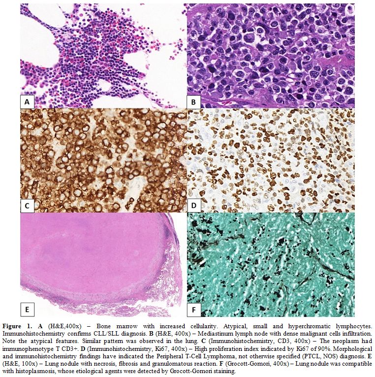
Figure 1. A (H&E,400x) – Bone marrow with increased cellularity. Atypical, small and hyperchromatic lymphocytes. Immunohistochemistry confirms CLL/SLL diagnosis. B (H&E, 400x) – Mediastinum lymph node with dense malignant cells infiltration. Note the atypical features. Similar pattern was observed in the lung. C (Immunohistochemistry, CD3, 400x) – The neoplasm had immunophenotype T CD3+. D (Immunohistochemistry, Ki67, 400x) – High proliferation index indicated by Ki67 of 90%. Morphological and immunohistochemistry findings have indicated the Peripheral T-Cell Lymphoma, not otherwise specified (PTCL, NOS) diagnosis. E (H&E, 100x) – Lung nodule with necrosis, fibrosis and granulomatous reaction. F (Grocott-Gomori, 400x) – Lung nodule was compatible with histoplasmosis, whose etiological agents were detected by Grocott-Gomori staining.  

Histoplasmosis is mostly found in immuno-compromised individuals, but can be found also in normal individuals; morphological diagnosis is accepted, but culture should confirm this etiology.[4]
CLL/SLL patients have an increased risk for development of a second neoplasm. Skin and lung cancer are the best examples. Another lymphoma as a second neoplasm, in spite of the possibility, is rarer (approximately 8%).[5] Tsimberidou et al.[6] reported a risk of a second neoplasm of 2.2 times higher than the expected risk in CLL/SLL patients at M.D. Anderson Cancer Center.[6]
Boyer et al.[7] reported three patients with CLL/SLL who have, in two cases, an ALK Anaplastic Large Cell Lymphoma (ALCL), and, in one case, a PTCL, NOS, as our patient.[5] Richter syndrome occurs in 5-10% and represents the transformation from low-grade to high-grade lymphoma. The most common is the transformation to Diffuse Large B-Cell Lymphoma (DLCBL). PTCL rarely happens in Richter Syndrome. Boyer et al.[7] studied three cases with other 21 patients reported in literature: all were elderly, with poor prognosis and their second neoplasm was diagnosed after some years of the first CLL/SLL diagnosis, as we observed in our patientl.[7]
There is not a well-understood theory about the association of PTCL and CLL/SLL. We believe that prior therapy may arise the risk due to immunosuppression, which is added to the cancer immunodeficiency by the CLL/SLL, with possible interactions between the drugs and T-cells.[7] In this context, Maddocks-Christianson et al.[7] found a significantly increased risk of second lymphoma in patients whose treatment included purine nucleoside analogues compared with patients who had not received those drugs. Besides, these authors did not find associations between the development of a second lymphoma with specific CLL/SLL patterns like ZAP-70, CD38, IgVH mutation status or cytogenetic abnormalities.[8]
Second neoplasms are also studied in the context of others lymphomas. Lung cancer and cutaneous melanoma risks are more elevated after CLL/SLL and Folicular Lymphoma (FL) than after DLBCL. Acute non-lymphocytic leukemia is more common after FL and DLBCL than after CLL/SLL.[8] So, the type of lymphoma influences the possibility of the second neoplasm.
Our case is uncommon and interesting. There is a high-grade lymphoma in a patient with a previous CLL/SLL under treatment, without residual disease. In the moment of the new diagnosis, he also has a reactivation of a fungi infection, probably by the immunosuppression of the second and aggressive lymphoma. Immunological alterations, previous treatment, genetic susceptibilities, infections agents are factors that may contribute for developing of second malignances. Clinics and pathologists must be alert for this sort of situation and due to this we send this letter. 


References      

  1. Beiggi S, Johnston JB, Seftel MD, Pitz MW, Kumar R, Banerji V, Griffith EJ, Gibson SB. Increased risk of second malignancies in chronic lymphocytic leukaemia patients as compared with follicular lymphoma patients: a Canadian population-based study. British Journal of Cancer. 2013;109:1287–1290. https://doi.org/10.1038/bjc.2013.381 PMid:23860531 PMCid:PMC3778273    
  2. Muzio M, Fonte E, Caligaris-Cappio F. Toll-like Receptors in Chronic Lymphocytic Leukemia. Mediterr J Hematol Infect Dis 2012, 4(1): e2012055. https://doi.org/10.4084/MJHID.2012.055    
  3. Aisenberg AC, Wilkes BM, Jacobson J. Rearrangement of the Genes for the Beta and Gamma Chains of the T Cell Receptor Is Rarely Observed in Adult B Cell Lymphoma and Chronic Lymphocytic Leukemia. J Clin Invest. 1987;80:1209-1214. https://doi.org/10.1172/JCI113182 PMid:2821076 PMCid:PMC442368    
  4. De D., Nath U. K. Disseminated Histoplasmosis in Immunocompetent Individuals- not a so Rare Entity, in India. Mediterr J Hematol Infect Dis 2015, 7(1): e2015028. https://doi.org/10.4084/MJHID.2015.028    
  5. Schöllkopf, Rosendahl D, Rostgaard K, Pipper C, Hjalgrim H. Risk of second cancer after chronic lymphocytic leukemia. Int. J. Cancer, 2007;121:151–156. https://doi.org/10.1002/ijc.22672 PMid:17351903      
  6. Tsimberidou AM, Wen S, McLaughlin O, O'Brien S, Wierda WG, Lerner S, Strom LS, Freireich EF, L, Medeiros J, Kantarjian HM, Keating MJ. Other Malignancies in Chronic Lymphocytic Leukemia/ Small Lymphocytic Lymphoma. J Clin Oncol.2009;27:904-910. https://doi.org/10.1200/JCO.2008.17.5398 PMid:19114699 PMCid:PMC4979239    
  7. Boyer DF, Lindeman NI, Harris NL, Ferry JA. Peripheral T-cell Lymphomas With Cytotoxic Phenotype in Patients With Chronic Lymphocytic Leukemia/Small Lymphocytic Lymphoma. Am J Surg Pathol.2014;38:279–288. https://doi.org/10.1097/PAS.0000000000000140 PMid:24418862      
  8. Maddocks-Christianson K, Slager SL, Zent CS, Reinalda M, TG Call, Habermann TM, Bowen A, Hoyer JD, Schwager S, Jelinek SF, E Neil and Shanafelt. Risk factors for development of a second lymphoid malignancy in patients with chronic lymphocytic leukaemia. British Journal of Haematology, 2007;139,398–404 https://doi.org/10.1111/j.1365-2141.2007.06801.x PMid:17910629    


[TOP]