The Impact of Chemotherapy after Pediatric Malignancy on Humoral Immunity to Vaccine-Preventable Diseases

Chiara Garonzi1, Rita Balter1, Gloria Tridello1, Anna Pegoraro1, Manuela Pegoraro2, Monia Pacenti3, Novella Scattolo4 and Simone Cesaro1.

 1 Pediatric Hematology Oncology, Azienda Ospedaliera Universitaria Integrata Verona, Italy.
2 Microbiology and Virology Unit, Azienda Ospedaliera Universitaria Integrata Verona, Italy.
3 Microbiology and Virology Unit, Padova University Hospital, Padova, Italy.
4 Ospedale di San Bonifacio, Azienda AULSS 9 Scaligera, Verona, Italy.

Corresponding author: Chiara Garonzi, M.D. Pediatric Hematology Oncology, Department of Mother and Child, Azienda Ospedaliera Universitaria Integrata Verona. Tel +39-045-8127163, fax: +39-045-8127830. E-mail: garonzi.chiara@gmail.com 

Published: March 1, 2020
Received: November 30, 2019
Accepted: February 9, 2020
Mediterr J Hematol Infect Dis 2020, 12(1): e2020014 DOI 10.4084/MJHID.2020.014

This is an Open Access article distributed under the terms of the Creative Commons Attribution License
(
https://creativecommons.org/licenses/by-nc/4.0), which permits unrestricted use, distribution, and reproduction in any medium, provided the original work is properly cited.

Abstract

Background/Aim: The antibody titer of vaccine-preventable disease in pediatric patients who underwent chemotherapy was assessed in order to evaluate the seroprotection after treatment and the feasibility and the efficacy of a policy of revaccination.
Methods: Serum antibody titers of 55 patients for hepatitis B (HBV), rubella, varicella-zoster (VZV), measles, mumps, polio viruses, Clostridium tetani (C. tetani) and Streptococcus pneumoniae (S. pneumoniae) were analysed.
Results: After chemotherapy, a lack of protective antibody titers against HBV, rubella, VZV, measles, mumps, polio viruses, C. tetani, and S. pneumoniae was found in 53%, 45%, 46%, 46%, 43%,  21-26%,  88% and 55% of patients, respectively. In 49 of 55 patients who were tested both before and after chemotherapy for at least a pathogen, the loss of immunity for HBV, rubella, VZV, measles, mumps, polio viruses and C. tetani was respectively 39%, 43%, 38%, 42%, 32%, 33%, and 80%. A low number of B-lymphocytes was associated with the loss of immunity against measles (p=0.04) whereas a high number of CD8+ T-lymphocytes was associated with the loss of immunity against VZV (p=0.03). A single booster of vaccine dose resulted in a seroprotection for HBV, rubella, VZV, measles, mumps, polio viruses, C. tetani and S. pneumoniae in 67%, 83%, 80%, 67%, 33%, 100%, 88% and 67% of patients, respectively.
Conclusions: We confirm that seroprotection for vaccine-preventable disease is affected by treatment for pediatric malignancy. A single booster dose of vaccine might be a practical way to restore vaccine immunity in patients after chemotherapy.


Introduction

Vaccination is one of the mayor conquests of public health.[1] It is fundamental in preventing several diseases, which could be life-threatening. Many positive results have been achieved so far, but global commitment should remain high in this field.
Patients with malignancies have an elevated risk of infections, some of them preventable by active immunization, e. g. invasive pneumococcal diseases.[2] This augmented susceptibility is related to the immune impairment due to the disease itself or the therapies (chemotherapy, radiotherapy).[3–5] Chemotherapy causes a transient immunodeficiency that lasts for about 6 months after the end of the treatment.[6] Moreover, in patients who undergo haemopoietic stem cell transplantation (HSCT), humoral and cellular immunity is affected by chemotherapy and radiotherapy used in the conditioning regimen, and by immunosuppressive treatment used to prevent graft rejection and graft versus host disease.[7]
International guidelines recommend revaccination from at least 3-6 months after the end of chemotherapy,[8] and from 3-6 to 12-24 months after HSCT.[9]
In this study, we assessed patients’ vaccination status at the diagnosis of malignancy, comparing it to that of a healthy population. Moreover, the persistence of protective antibody titer was followed-up after the end of chemotherapy in order to apply a re-vaccination protocol and assess its efficacy in restoring vaccination immunity. 

Material and Methods

Patients. Two hundred thirty-nine patients with a diagnosis of malignancy from 2010 to October 2017 were evaluated for inclusion in the study. Eligibility criteria were patients with age < 18 years, a previous diagnosis of malignancy and being off from chemotherapy for at least 6 months. Patients who undergo HSCT were excluded from the study (n=15). The period of recruitment was from January to June 2018. To exclude the potential bias related to different schedules of vaccination or different access to vaccination programs according to the country of origin, the authors  recruited only Italian patients or patients born in Italy.  The final study group comprised of 55 patients. Clinical data and antibody titers at the diagnosis were obtained retrospectively, consulting electronic medical records. As part of an internal program of revaccination, each eligible patient was assessed for serologic antibodies against hepatitis B (HBV), rubella, varicella-zoster (VZV), measles, mumps, polio viruses, Clostridium tetani (C. tetani) and Streptococcus pneumoniae (S. pneumoniae), and, at the same time, the immune recovery was evaluated by counting the number of CD3+, CD4+, CD8+ and CD19+ lymphocytes. Informed consent was obtained from parents for blood sampling needed to determine the serological status for vaccine-preventable diseases after chemotherapy. Patients who were not protected against at least one disease and who were eligible (n=46) were invited to perform revaccination according to internal guidelines for vaccinations that are shown in table 1. Twenty-one of 46 patients (46%) were compliant with the revaccination protocol. In 10 of 21 patients (48%), the efficacy of revaccination was assessed after at least one month after immunization. Data collection was closed as of January 31st, 2019. 

Table 1 
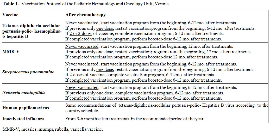
Table 1. Vaccination Protocol of the Pediatric Hematology and Oncology Unit, Verona.

Serologic assays. Antibodies detection for HBV, rubella, VZV, measles, mumps, polio viruses, C. tetani, and S. pneumoniae were conducted on fresh serum samples using commercial kits and following the manufacturer’s instructions. Chemiluminescence technology (CLIA) was used to detect rubella, measles, mumps, VZV IgG (Diasorin S.p.A., Saluggia, Vercelli, Italy) and HBsAg antibodies (Siemens Healthcare Diagnostics, Inc., Tarrytown, NY, US); enzyme immuno assay (EIA) was used for C. tetani IgG (Diasorin S.p.A., Saluggia, Vercelli, Italy) and for S. pneumoniae IgG (The Binding Site Group Ltd, Birmingham, UK); neutralization assay, according to WHO guidelines, was used for poliovirus. Results were interpreted as positive, negative or undetermined according to kit instructions. Positive results were considered as follow: HbsAg antibodies > 12 mUI/ml, rubella IgG > 10 UI/mL, VZV IgG > 135 mUI/mL, measles IgG > 16,5 AU/mL, mumps IgG > 9 AU/mL, C. tetani IgG > 0,51 UI/mL, poliovirus IgG > 1:8. For S. pneumoniae were considered positive results IgG titers >35 mg/L, in accordance with WHO recommendation.[10] Specifically, the kit used measures antibody responses to pneumococcal vaccines incorporating 23 polysaccharides isolated from S. pneumoniae.
Statistical analysis. Collected data were analyzed using descriptive statistics: median and range for continuous variables, absolute frequency, and percentages for categorical or dichotomous variables.
Differences of quantitative variables between groups were texted using the non-parametric Kruskal-Wallis test, whereas categorical variables using the Chi-squared test or Fisher’s exact test.
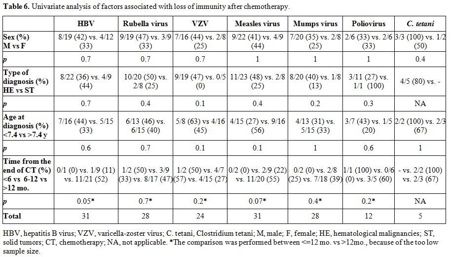
The following variables were tested in univariate analysis for the loss of protective serum antibody titers: sex, type of diagnosis, age at diagnosis, time from the end of chemotherapy.
A P-value of <0,05 was considered statistically significant. Analysis was conducted using the statistical software SAS, 9.4 version (Statistical Analysis Software, SAS Institute Inc.).
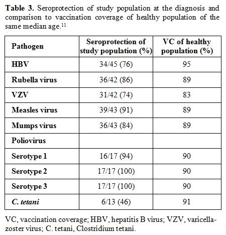
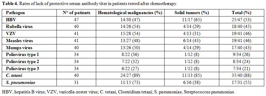
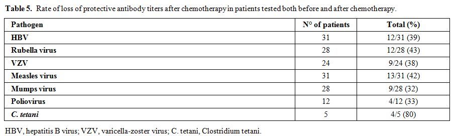
The term lack of immunity was used for patients with non-protective antibody titers if they were tested after chemotherapy. The term loss of immunity was used for patients tested pre- and post- treatment and had a change of the antibody level from positive to negative. Seroprotection was defined by any level of antibody positivity, including a borderline positivity with an undetermined titer.

Results

The main demographic and clinical characteristics of patients are shown in table 2.

Table 2 Table 2. Demographic and clinical characteristics of study patients.

Antibody titers at diagnosis. At diagnosis, the complete serological panel of antibodies against HBV, rubella, VZV, measles, mumps, polio viruses, and C. tetani was available only for 11 of 55 patients, whereas 4 patients did not perform any antibody determination. The most missing data were antibodies against poliovirus (available only for 22 patients) and C. tetani (available for 15 patients). In table 3, data of seroprotection in the study population at the diagnosis (median age: 5.9 years) were compared to regional vaccination coverage (VC) in a healthy population of at the latest 8 years old.[11]

Table 3 Table 3. Seroprotection of study population at the diagnosis and comparison to vaccination coverage of healthy population of the same median age.[11]

Some patients lack protective immunity at diagnosis, in detail, 11 patients (24%) for HBV, 6 (14%) for rubella virus, 11 (26%) for VZV, 4 (9%) for measles virus, 7 (16%) for mumps virus, 1 (6%) for poliovirus and 7 (54%) for C. tetani. The lack of seroprotection was related to the fact that patients were not vaccinated in 1/11 for HBV, 2/6 for rubella virus, 5/11 for VZV, 2/4 for measles virus, and 2/7 for the mumps virus.
Lack of protective serum antibody titers after chemotherapy. Fifty-five patients assessed the antibody titers after a median time of 26 months from the end of chemotherapy. The rate of the lack of protective serum antibody titers for HBV, rubella, VZV, measles, mumps, polio viruses, C. tetani and S. pneumoniae is presented in table 4. Data differ by type of vaccine and underlying malignancy, ranging from 27-36% for poliovirus to 89% for C. tetani in the group with hematological malignancies and from 8% for poliovirus to 85% of patients for C. tetani in solid tumors group.


Table 4 Table 4. Rates of lack of protective serum antibody titer in patients tested after chemotherapy.

Loss of protective serum antibody titers after chemotherapy. In patients tested both before and after chemotherapy, rates of protective antibody titers loss for HBV, rubella, VZV, measles, mumps, polio viruses, and C. tetani are summarized in table 5. Rates vary depending on vaccine type: from 32% of patients for mumps virus to 80% for C. tetani.

Table 5 Table 5. Rate of loss of protective antibody titers after chemotherapy in patients tested both before and after chemotherapy.

Univariate analysis for the loss of protective serum antibody titer is given in table 6. No factor was statistically significant.

Table 6 Table 6. Univariate analysis of factors associated with loss of immunity after chemotherapy.

The comparison between the number of CD3+, CD4+, CD8+ and CD19+ lymphocytes determined after chemotherapy and average values for age, i.e., 5-10 years old,[12] is shown in table 7.

Table 7 Table 7. Comparison between lymphocyte subpopulations in peripheral blood of study patients after chemotherapy and healthy controls.[12]

In univariate analysis, a lower number of B-lymphocytes resulted significantly associated with the loss of protective antibody titer for measles virus, p=0.04, whereas a higher number of CD8+ T-lymphocytes was significantly associated with the loss of antibody protection for VZV, p=0.03.
Antibody titers after vaccination protocol application. Revaccination with a single-shot vaccination boost of 10 patients who underwent chemotherapy obtained the restoration of protective serum antibody level in 2 out of 3 patients for HBV, in 5 out of 6 patients for rubella virus, in 4 out of 5 patients for VZV, in 4 out of 6 patients for measles virus, in 2 out of 6 patients for mumps virus, in 8 out of 8 patients for poliovirus, in 7 out of 8 patients for C. tetani and in 2 out of 3 patients for S. pneumoniae.

Discussion

This study was part of an internal program to stimulate adherence to the revaccination protocol after treatment with chemotherapy for malignancy. The assessment of vaccination serological coverage at the diagnosis showed that several patients were not protected. In twelve out of 47 (25%) seronegative status was attributable to the fact that patients were not compliant with the national vaccination program for newborn and child because of the young age at the diagnosis of malignancy or of parental decision. This last reason was observed especially for attenuated live virus vaccine where the viral replication by vaccine strain is perceived by parents as a potential risk for recurrence of the underlying disease. On the other hand, 33 out of 47 determinations (70%) resulted in negative despite that patients were vaccinated. It may be explained by the fact that some patients did not have completed the primary cycle of vaccination by the time of the diagnosis or the vaccination was ineffective.
In this study, data of protective immunity due to vaccination were not available for every patient, especially for C. tetani and poliovirus. This reflects a local problem because these testes were not performed at the hospital laboratory, and, if requested by the physician in charge, the serum samples had to be sent to laboratories outside, increasing the costs.
When the rate of patient seroprotection at the diagnosis was compared to the vaccination coverage in a healthy population, significant differences were seen for HBV (respectively 76% vs. 95%), poliovirus (94-100% vs. 90%), and C. tetani (46% vs. 91%). We underline that seroprotection figures in the study patients were calculated on the basis of antibody determinations, whereas in the general population, this index is based just on the administration of vaccination, irrespective of efficacy in terms of seroprotection. Therefore, the data on regional vaccination coverage for vaccines could not correspond to the efficacy of vaccination, intended as the documentation of a protective antibody titer after vaccination. Moreover, the differences could also be attributed to the small sample size. Considering the ideal immunization coverage recommended by World Health Organization in the health population to provide herd immunity, i.e., 95%, it emerged that patients’ immunization coverage was far from this value, except from polio (95-100%).
One of our principal purposes was to analyze the impact of chemotherapy on vaccine humoral immunity. We demonstrated that patients were prone to loose protective antibody titer, acquired by vaccination, or in some cases by natural infection. As a result, the patients after chemotherapy are frequently not protected against vaccine-preventable diseases, and this fact can be relevant when the patients return to school or in the community. In the case of outbreaks, like the measles outbreak which affected Italy in 2017,[13] these patients would be hugely at risk.
In literature, the loss of protective antibody titers after chemotherapy is frequent, even if percentages of loss or lack of immunity vary depending on the study considered, ranging from 25-88% for measles virus, 26-88% for mumps virus, 19-88% for rubella virus,[14–16] 17-35% for VZV,[14,17] 11.6-14% for C. tetani,[15,18] 7% for poliovirus,[15] and 26-86% for HBV.[15,16,19,20] In patients with Acute Lymphoblastic Leukemia, non-protective antibody levels were seen in 2-80% patients for C. tetani, 0-38% for poliovirus, 8-71% for mumps virus, 40-71% for measles virus and 8-28% for rubella virus.[21] Although these studies are heterogeneous, most of them were performed with patients with hematological malignancies, whereas our study included both hematological and solid tumors.
The risk factor analysis for the loss of immunity did not find any association with sex, type of diagnosis, age at diagnosis, and time from the end of chemotherapy, although we highlight that the sample was small. Interestingly, we showed that a low number of CD19+ lymphocytes was associated with the loss of protective antibody titer against measles virus. This observation is in line with the finding that patients treated with B-depleting monoclonal antibodies, such as rituximab, have lower responses to vaccination for at least 6 months after the end of treatment.[22,23] We also found that a high number of CD8+ lymphocytes is associated with the loss of immunity against VZV. This finding needs to be confirmed further.
In order to evaluate the role of the immune recovery in the persistence or the loss of vaccine immunity after therapies, the peripheral blood subpopulations of lymphocytes were assessed, and no differences were found between study patients and healthy children of the same age. We hypothesize that the re-exposure to vaccine antigens has a crucial role in vaccine immunity reconstitution in order to induce the appearance and proliferation of lymphocytes capable of mounting a specific humoral response again. For this reason, our policy was to revaccinate the patients starting from 6 months from the end of treatments. Forty-six percent of patients were compliant with this policy by the study period and this measure was associated with a seroprotection in 33-100% of patients after a single booster dose of vaccine.

Conclusions

This study confirms that the loss of humoral protection from vaccine preventable disease is a common finding among hematological and oncological patients after chemotherapy. The practice to revaccinate the patients to re-establish individual protection and to contribute to the herd immunity has a favorable benefit/risk ratio especially for the return to community activities or for travelling. Several important issues remain to define such as the optimal timing for revaccination (6-12 months vs. other) and the type of schedule (boost vs. full repeated doses) that requires further prospective studies on a larger number of patients. 
 

References   

  1. Centers for Disease Control and Prevention. Ten Great Public Health Achievements - United States, 1900-1999. MMWR Morb Mortal Wkly Rep. 1999;48:241-3.
  2. Meisel R, Toschke AM, Heiligensetzer C, Dilloo D, Laws HJ, Von Kries R. Increased risk for invasive pneumococcal diseases in children with acute lymphoblastic leukaemia. Br J Haematol. 2007;137:457-60. https://doi.org/10.1111/j.1365-2141.2007.06601.x PMid:17488489
  3. Friman V, Winqvist O, Blimark C, Langerbeins P, Chapel H, Dhalla F. Secondary immunodeficiency in lymphoproliferative malignancies. Hematol Oncol. 2016;34:121-32. https://doi.org/10.1002/hon.2323 PMid:27402426
  4. Chinen J, Shearer WT. Secondary immunodeficiencies, including HIV infection. J Allergy Clin Immunol. 2010;125:S195-203. https://doi.org/10.1016/j.jaci.2009.08.040 PMid:20042227 PMCid:PMC6151868
  5. Goswami M, Prince G, Biancotto A, Moir S, Kardava L, Santich BH, Cheung F, Kotliarov Y, Chen J, Shi R, Zhou H, Golding H, Manischewitz J, King L, Kunz LM, Noonan K, Borrello IM, Smith BD, Hourigan CS. Impaired B cell immunity in acute myeloid leukemia patients after chemotherapy. J Transl Med. 2017;15:155. https://doi.org/10.1186/s12967-017-1252-2 PMid:28693586 PMCid:PMC5504716
  6. Ek T, Mellander L, Andersson B, Abrahamsson J. Immune Reconstitution After Childhood Acute Lymphoblastic Leukemia Is Most Severely Affected in the High Risk Group. Pediatr Blood Cancer. 2005;44(5):461-8. https://doi.org/10.1002/pbc.20255 PMid:15558707
  7. Tomblyn M, Chiller T, Einsele H, Gress R, Sepkowitz K, Storek J, Wingard JR, Young JAH, Boeckh MJ. Guidelines for Preventing Infectious Complications among Hematopoietic Cell Transplantation Recipients: A Global Perspective. Biol Blood Marrow Transplant. 2009;15:1143-238. https://doi.org/10.1016/j.bbmt.2009.06.019 PMid:19747629 PMCid:PMC3103296
  8. Mikulska M, Cesaro S, de Lavallade H, Di Blasi R, Einarsdottir S, Gallo G, Rieger C, Engelhard D, Lehrnbecher T, Ljungman P, Cordonnier C, European Conference on Infections in Leukaemia group. Vaccination of patients with haematological malignancies who did not have transplantations: guidelines from the 2017 European Conference on Infections in Leukaemia (ECIL 7). Lancet Infect Dis. 2019;19:e188-99. Epub 2019 Feb 8. https://doi.org/10.1016/S1473-3099(18)30601-7
  9. Cordonnier C, Einarsdottir S, Cesaro S, Di Blasi R, Mikulska M, Rieger C, de Lavallade H, Gallo G, Lehrnbecher T, Engelhard D, Ljungman P, European Conference on Infections in Leukaemia group. Vaccination of haemopoietic stem cell transplant recipients: guidelines of the 2017 European Conference on Infections in Leukaemia (ECIL 7). Lancet Infect Dis. 2019;19:e200-12. Epub 2019 Feb 8. https://doi.org/10.1016/S1473-3099(18)30600-5
  10. Ciofi degli Atti ML, Azzari C, Bartolozzi G, Esposito S, Fara GM, Giovanetti F, Lo Giudice M. I correlati immunologici di protezione indotti dalle vaccinazioni. Rivista di Immunologia e Allergologia Pediatrica. 2010;1:27-31. Available from: https://www.siaip.it/upload/riap/327_I%20correlati%20immunologici%20di%20protezione%20indotti%20dalle%20vaccinazioni.pdf  
  11. Ministero della Salute. Vaccinazioni dell'età pediatrica. Anno 2017 (coorte 2009). Coperture vaccinali (per 100 abitanti) per vaccinazioni eseguite entro gli 8 anni, calcolate sui riepiloghi inviati dalle Regioni e PP.AA. (per singolo antigene) [Internet]. [cited 2019 Jul 17]. Available from: http://www.salute.gov.it/imgs/C_17_tavole_20_allegati_iitemAllegati_4_fileAllegati_itemFile_6_file.pdf
  12. Fiore M, Duse M. Guida pratica. Le immunodeficienze nell'ambulatorio del pediatra [Internet]. 2016 [cited 2019 Jul 17]. Available from: https://www.aip-it.org/sites/default/files/allegati-pagine/guidaimmunodeficienze.pdf
  13. Filia A, Bella A, Del Manso M, Baggieri M, Magurano F, Rota MC. Ongoing outbreak with well over 4,000 measles cases in Italy from January to end August 2017 − what is making elimination so difficult? Euro Surveill. 2017;22. https://doi.org/10.2807/1560-7917.ES.2017.22.37.30614 PMid:28933342 PMCid:PMC5607657
  14. Bochennek K, Allwinn R, Langer R, Becker M, Keppler OT, Klingebiel T, Lehrnbecher T. Differential loss of humoral immunity against measles, mumps, rubella and varicella-zoster virus in children treated for cancer. Vaccine. 2014;32:3357-61. https://doi.org/10.1016/j.vaccine.2014.04.042 PMid:24793952
  15. Zignol M, Peracchi M, Tridello G, Pillon M, Fregonese F, D'Elia R, Zanesco L, Cesaro S. Assessment of humoral immunity to poliomyelitis, tetanus, hepatitis B, measles, rubella, and mumps in children after chemotherapy. Cancer. 2004;101:635-41. https://doi.org/10.1002/cncr.20384 PMid:15274078
  16. Keskin Yildirim Z, Buyukavci M. Assessment of Humoral Immunity to Hepatitis B, Measles, Rubella, and Mumps in Children After Chemotherapy. J Pediatr Hematol Oncol. 2018;40:e99-102. https://doi.org/10.1097/MPH.0000000000001072 PMid:29309372
  17. Patel SR, Bate J, Maple PA, Brown K, Breuer J, Heath PT. Varicella zoster immune status in children treated for acute leukemia. Pediatr Blood Cancer. 2014;61:2077-9. https://doi.org/10.1002/pbc.25086 PMid:24789692
  18. Kwon HJ, Lee JW, Chung NG, Cho B, Kim HK, Kang JH. Assessment of serologic immunity to diphtheria-tetanus- pertussis after treatment of Korean pediatric hematology and oncology patients. J Korean Med Sci. 2012;27:78. https://doi.org/10.3346/jkms.2012.27.1.78 PMid:22219618 PMCid:PMC3247779
  19. Fayea NY, Kandil SM, Boujettif K, Fouda AE. Assessment of hepatitis B virus antibody titers in childhood cancer survivors. Eur J Pediatr. 2017;176:1269-73. https://doi.org/10.1007/s00431-017-2970-4 PMid:28730317
  20. Karaman S, Vural S, Yildirmak Y, Urganci N, Usta M. Assessment of hepatitis B immunization status after antineoplastic therapy in children with cancer. Ann Saudi Med. 2011;31:573-6. https://doi.org/10.4103/0256-4947.87091 PMid:22048500 PMCid:PMC3221126
  21. van Tilburg CM, Sanders EA, Rovers MM, Wolfs TF, Bierings MB. Loss of antibodies and response to (re-)vaccination in children after treatment for acute lymphocytic leukemia: a systematic review. Leukemia. 2006;20:1717-22. https://doi.org/10.1038/sj.leu.2404326 PMid:16888619
  22. van der Kolk LE, Baars JW, Prins MH, van Oers MHJ. Rituximab treatment results in impaired secondary humoral immune responsiveness. Blood. 2002;100:2257-9. https://doi.org/10.1182/blood.V100.6.2257 PMid:12200395
  23. Hua C, Barnetche T, Combe B, Morel J. Effect of methotrexate, anti-tumor necrosis factor α, and rituximab on the immune response to influenza and pneumococcal vaccines in patients with rheumatoid arthritis: A systematic review and meta-analysis. Arthritis Care Res. 2014;66:1016-26. https://doi.org/10.1002/acr.22246 PMid:24339395

Supplementary Tables:

Suppl Table 1 Supplementary table 1. Univariate analysis of the number of lymphocytes in peripheral blood and the loss of protective antibody titers after chemotherapy.

Suppl Table 2 Supplementary table 2. Presence of protective antibody titers after application of Vaccination Protocol, after chemotherapy.

Suppl Table 3 Supplementary table 3. Vaccine immunity reconstitution in patients with lack of immunity after chemotherapy, after the application of Vaccination Protocol. 


[TOP]