Epidemiology and Clinical Characteristics of Henoch-Schönlein Purpura Associated with Epstein-Barr Virus Infection

Hong-bo Hu1, Jian-gang Wu2, Ying Cheng3 and Jian-jun Li4..

1Department of Laboratory, Maternal and Child Health Hospital of Hubei Province, China.
2 Department of Infectious Disease, The first people's Hospital of Guangshui, China
3 Department of Pediatrics, Maternal and Child Health Hospital of Hubei Province, China
4 Medical department, The first people's Hospital of Guangshui, China.

Correspondence to: Jian-jun Li, MD, The first people’s Hospital of Guangshui, Guangshui, Hubei 432700, China. E-mail: jianjunlgs1@163.com  

Published: November 1, 2021
Received: July 26, 2021
Accepted: October 16, 2021
Mediterr J Hematol Infect Dis 2021, 13(1): e2021064 DOI 10.4084/MJHID.2021.064

This is an Open Access article distributed under the terms of the Creative Commons Attribution License
(
https://creativecommons.org/licenses/by-nc/4.0), which permits unrestricted use, distribution, and reproduction in any medium, provided the original work is properly cited.

Abstract

Background: Henoch-Schönlein purpura (HSP) is an immune-mediated vasculitis, and the formation of immune complexes may be triggered by exposure to Epstein-Barr virus (EBV) infection.

Methods: We performed a five-year case-control study to evaluate the epidemiology and clinical characteristics of HSP associated with EBV infection.

Results: The incidence of EBV-triggered HSP was 4.2%, while EBV infection in children with HSP was 0.9%; The EBV-triggered HSP cases had a significantly higher frequency of abdominal pain than the Mycoplasma Pneumoniae (MP)-triggered HSP group (χ2 = 8.024, p = 0.005); Significant differences were observed in the duration of abdominal pain (Z = -1.935, p = 0.027) between the two groups; C3 (t = 9.709, p < 0.001), IgA (t = 20.39, p < 0.001) and IgG (t = 6.407, p < 0.001) were significantly increased in the EBV infection group than those in the healthy control group. Notably, significantly higher proportion of CD19 (t = 6.773, p < 0.001) and lower proportion of CD56 (t = 11.13, p < 0.001) was found in EBV infection group compared with healthy control group. The IgA level was higher than that of the non-infectious group (t = 2.162, p = 0.032), but their CD4/CD8 ratio (t = 10.070, p < 0.001) and CD56 proportion (t = 2.096, p = 0.037) were significantly lower.

Conclusions: Both cellular and humoral immunity were involved in the pathogenesis of EBV-triggered HSP, leading to increased production of inflammatory mediators and immunoglobulins. Those events may cause or promote the development of systemic vessel vasculitis.


Introduction

Epstein-Barr virus (EBV) belongs to the gamma subfamily of herpesviruses and is classified as human herpesvirus 4. It is the main and important pathogen in children worldwide that seriously threatens their health and burdens patients, families, and society.[1,2]
It has been reported that EBV is associated with various forms of vasculitis, such as Kawasaki disease, rheumatoid vasculitis, central nervous system vasculitis, and cutaneous necrotizing vasculitis.[3-6] Henoch-Schönlein purpura (HSP) is the most common vasculitis in children characterized by palpable purpura, arthritis, gastrointestinal symptoms, and renal involvement. Infections may be one of the several possible triggers participating in its pathogenesis.[7,8] EBV has also been reported to initiate HSP,[9-11] but these reports are mostly individual cases. The data on the etiology and epidemiology of children with HSP and EBV infection are still insufficient.
This study investigated the incidence of EBV-triggered HSP. Further, it estimated the epidemiological characteristics of affected children, such as their age, gender, seasonal distribution, clinical manifestations, and related laboratory findings, to provide researchers and clinicians with more clinical data, which may help study EBV infection's role in the etiology of HSP.

Methods

Patient selection. We recruited all patients with HSP between the ages of 2 and 15 years who were admitted to two hospitals in Hubei province, China, from January 2015 to December 2019. The diagnosis of HSP was based on the European League against Rheumatism endorsed consensus criteria for HSP classification.[12] Children with HSP, excluding those with infection as a trigger, served as non-infectious cases for direct comparison. MP-triggered HSP cases, firstly reported in another paper,[7] were also included in the control groups.
Laboratory tests for EBV infection
. Blood samples from patients were tested for anti-EBV viral capsid antigen (anti-EBV VCA- IgG), EBV capsid antigen (anti-EBV VCA-IgM), early antigen (anti-EBV EA-IgG) antibodies, or/and EBV-DNA. The diagnosis of an acute EBV infection was determined by one or more of the following criteria: (1) detection of anti-EBV VCA-IgM; (2) transient antibody response to anti-EBV EA-IgG; and (3) virus DNA concentration was ≥ 1 × 10³ copies/mL in whole blood which was measured by real-time fluorescent quantitative PCR.[13] In addition, children diagnosed with EBV infection, regardless of HSP, were surveyed.
Laboratory data collection
. The patients underwent other laboratory tests, such as cluster of differentiation (CD)3, CD4, CD8, CD19, CD56, immunoglobulin (Ig)A, IgM, IgG, complement (C) 3, and C4 concentrations. The differences in the laboratory findings among groups were studied by matched-pair analyses (including healthy and MP-triggered HSP controls). For each EBV-triggered HSP case, control groups were selected, which were matched for sex, age, and admission time.
Exclusion criteria
. The exclusion criteria were as follows: (1) patients with impaired immune functions or those who were receiving immunosuppressive therapies or were taking nephrotoxic drugs; (2) patients who received blood transfusions or other blood product treatment in the past six months; (3) patients with severe heart, liver, kidney, or other organ system diseases; (4) patients with incomplete clinical data; (5) patients with HSP associated with infections caused by other pathogens.
Statistical analyses. Statistical analyses were performed using the SPSS ver. 21.0 software (SPSS, Inc., Chicago, IL, USA). Comparisons of the frequencies among groups were analyzed using Chi-square tests. In cases in which the data were not normally distributed, the nonparametric Mann-Whitney U test was applied. Comparisons of mean values between groups were performed using the independent sample t-tests. A P-value of < 0.05 was considered statistically significant.

Results

Incidence of EBV-triggered HSP. A total of 1437 children diagnosed with HSP were enrolled; among them, 819 were male and 618 were female, with a male-to-female ratio of 1.33:1. The 61 HSP children with EBV infection included 38 boys and 23 girls (male: female = 1.65:1). The incidence of EBV-triggered HSP was 4.2% (61/1437).
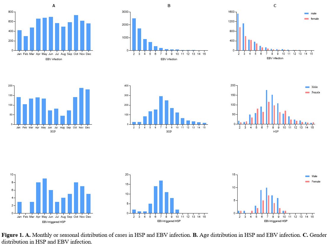
Monthly of cases of HSP and EBV infection
. The incidence of HSP cases was lower in summer and higher in winter, while EBV infection and EBV-triggered HSP had no obvious seasonal characteristics (Figure 1A).
Age and gender distribution of HSP and EBV infection
. A peak prevalence, mainly in the 2–5 years age range, was observed in the EBV infection cases, and after this age period, the prevalence decreased dramatically. However, a bell-shaped distribution pattern was also seen with a peak prevalence mainly in the 6–10 years age range in the HSP and EBV-triggered HSP cases (Figure 1B). There was no significant difference in the male-to-female ratio in the three groups (χ2 = 0.418, p = 0.811, Figure 1C).


Figure 1 Figure 1. A. Monthly or seasonal distribution of cases in HSP and EBV infection. B. Age distribution in HSP and EBV infection. C. Gender distribution in HSP and EBV infection.

Clinical manifestations of EBV infection. Six thousand seven hundred ninety-four children were determined with positive results, including 4098 males and 2696 females (male: female=1.52;1). A total of 63807 children were tested for EBV, and the rate of EBV infection was 10.6% (6794/63807) (Table 1). The frequency of EBV infection developing HSP in children was 0.9% (61/6794). In the cases of EBV infection, a significantly greater frequency of EBV-triggered HSP was found in the age of 6-10 years (χ2 = 356.808, p < 0.001).

Table 1 
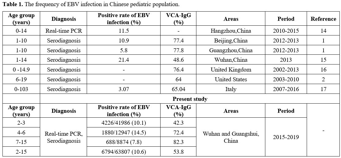
Table 1. The frequency of EBV infection in Chinese pediatric population.

Clinical manifestations of HSP with EB infection. The EBV-triggered HSP cases most frequently exhibited abdominal pain (39/61, 63.9%), followed by arthritis/arthralgia (34/61, 55.7%), and renal involvement (12/61, 20.0%) (Table 2). The clinical manifestations presented no significant heterogenicity between the EBV-triggered HSP group and the non-infectious group. However, the EBV-triggered HSP cases had a significantly higher frequency of abdominal pain than the MP-triggered HSP group (χ2 = 8.024, p = 0.005) (Table 3).

Table 2 
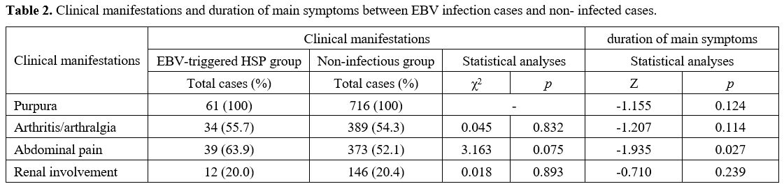
Table 2. Clinical manifestations and duration of main symptoms between EBV infection cases and non- infected cases. 

Table 3 
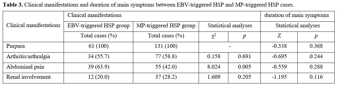
Table 3. Clinical manifestations and duration of main symptoms between EBV-triggered HSP and MP-triggered HSP cases.

Duration of main symptoms. The comparison of the duration of main symptoms of the non-infectious cases with infectious cases is presented in Table 2. Significant differences were observed in the duration of abdominal pain (Z = -1.935, p = 0.027) between the two groups.
Laboratory tests
. As shown in Figure 2, C3 (t = 9.709, 
p < 0.001), IgA (t = 20.39, p < 0.001) and IgG (t = 6.407, p < 0.001) were significantly higher in the EBV-infected group than those in the healthy control group. Notably, a significantly higher proportion of CD19 (t = 6.773, p < 0.001) and lower proportion of CD56 (t = 11.13, p < 0.001) was also found in EBV-infected group compared with healthy control group.
The IgA level of the infected group was also significantly higher than that of the non- infected group (t = 2.162, 
p = 0.032), while CD4/CD8 ratio (t = 10.070, p < 0.001) and CD56 proportion (t = 2.096, p = 0.037) were significantly lower than those of the non- infected group.

Figure 2 
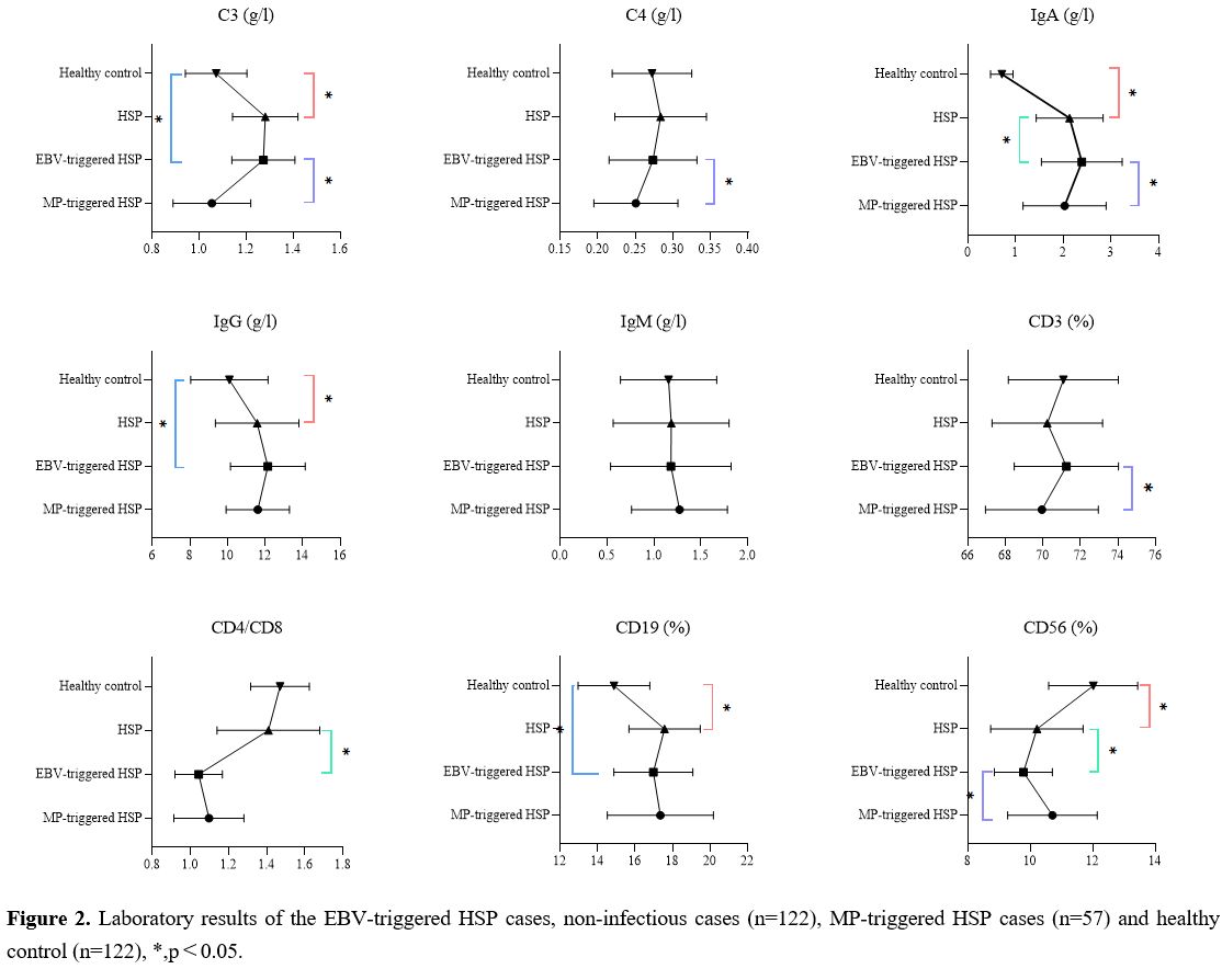
Figure 2.  Laboratory results of the EBV-triggered HSP cases, non-infectious cases (n=122), MP-triggered HSP cases (n=57) and healthy control (n=122), *, p<0.05.

The laboratory results of C3 (t = 7.926, p < 0.001), C4 (t = 2.168, p = 0.032), IgA (t = 2.278, p = 0.025) and CD3 proportion (t = 2.450, p = 0.016) were significantly decreased in the EBV infection group than in the MP-triggered HSP group, while the CD56 proportion (t =4.228, p < 0.001) increased significantly.

Discussion

EBV seropositivity among populations has much geographic variation.[1,2,14-17] Our study showed that the EBV seroprevalence rate of Chinese children aged 2-3 years was 42.3%, and about 82.3% of children aged 7-15 years had serological evidence of EBV infection. These percentages are similar to other reports on Chinese patients.[1,14,15] In recent studies, the frequency of EBV-triggered HSP has been reported as 7.2% to 8.5%, and the incidence of HSP cases in EBV infection was approximately 0.5-3.9% in China.[18-21] In our study, the incidence of EBV infection in HSP cases was 4.2%, while that of EBV infection developing HSP in children was 0.9%. To our knowledge, only minimal information about the prevalence of EBV-triggered HSP is available in other countries and regions. Multi-centric studies with larger sample sizes from diverse backgrounds may provide more information about EBV-triggered HSP.
The seasonality of HSP is determined by the infection that triggers it.[7,22] However, no clear seasonal changes in the distribution of EBV-positive cases were found each month. Thus, the season might not be an influencing factor of EBV infection.
The gender distribution of the incidence of HSP was also reported in some studies.[4,23,24] In this study, 819 were male, 618 were female, with a male-to-female ratio of 1.33:1. Based on the results, the boys were affected more often than the girls, and these findings were similar to earlier reports.[8,23,24] Thus, the gender distribution of EBV infection was similar to that of HSP.
Our findings demonstrated that EBV infection peaked in patients aged 2–5 years. However, a significantly greater frequency of EBV-triggered HSP was found in 6-10 years. HSP is a common immune-mediated disease in children characterized by leukocytoclastic vasculitis involving the capillaries and IgA immune complexes' deposition. The entire IgA system is naturally deficient at the preschool stage, and it is manifested by the deficiency or low level of serum IgA and paucity of IgA-producing cells in various tissues. As a marker of IgA system maturation the serum IgA level reaches the adult in adolescence,[25] and that is probably one of the reasons why EBV-triggered HSP mainly occurs at school-age.
We have done an investigation on the clinical manifestations of MP-triggered HSP in previous studies.[11] The EBV-triggered HSP cases had a higher frequency of abdominal pain than the MP-triggered HSP group, and a significantly longer duration of abdominal pain was found in the EBV infection group compared with the non-infectious group. Wang et al. also concluded that EBV infection may be a risk factor for refractory gastrointestinal HSP after investigating the clinical outcome of mycophenolate mofetil in pediatric refractory gastrointestinal HSP.[9]
Studies about the EBV-induced differentiation of B-cells into IgA-secreting cells suggested a possible involvement of EBV in the pathogenesis of the systemic immune-mediated disease. It has been demonstrated that patients with systemic lupus erythematosus had strongly increased IgA levels against the EBV antigens compared to healthy individuals.[26] Similarly, the IgA levels were also significantly increased in the EBV-triggered HSP group than in the healthy control and non-infectious groups. These findings may support the association between EBV infection and HSP.
It has been demonstrated that the number of CD4+T cells and the CD4/CD8 ratio were reduced, while the number of CD8+T lymphocytes was increased in patients with HSP.[27] In the present study, the CD4/CD8 ratio was lower than that of HSP. During EBV infection, B‐cells are infected first, followed by the activation of CD8+ T‐cells. The TNF-α secreted by the activated CD8+ T cells can promote the release of other cytokines and chemokines, leading to tissue damage, stimulating endothelial cell and mesangial cell mitosis, and causing vascular inflammation synergistically with IgA.[27] Consequently, we considered that EBV infection might lead to or aggravate the immune dysfunction of patients.
Natural killer (NK) cells play a vital role in the protective immunity against pathogen-infected cells. However, NK cell regulation disorder may hinder the clearance of exogenous antigens, resulting in immune damage. Consistent with previous studies,[28] we found that the proportion of CD56 in peripheral blood of HSP patients decreased, and this proportion was even lower in EBV-triggered HSP cases. Similar findings were found in a study on EBV-triggered juvenile dermatomyositis.[29] Interestingly, comparing EBV-triggered HSP patients with those MP-triggered, laboratory tests showed a significant increase in the proportion of CD56 in the latter group. The previous study has implied that NK cells are over-activated and proliferated in Mycoplasma pneumoniae pneumonia cases, and the activation of NK cells may be indispensable in the pathogenesis of pneumonia. Therefore, more attention should be given to the role of NK cells in the pathogenesis of EBV-triggered HSP.
Although both EBV and MP infection may trigger HSP, our laboratory results show that the pathogenesis of HSP caused by these two pathogens may not be the same. Compared with the EBV-triggered HSP group, our results proved that IgA, C3, C4 and CD3 were significantly decreased in MP-triggered HSP patients. HSP has been confirmed to be related to the activation of the complement system, while complement can mediate MP cleavage through classical and alternative pathways and trigger an inflammatory response.[30-32] Since C3 and C4 contribute to promoting the body's synthesis of antibodies, excessive consumption of complement components will eventually reduce the body's antibody levels. Therefore, we inferred that MP-triggered HSP might be caused by complement activation due to MP infection. Further study on these differences is required.


Conclusions

Herein, we present 61 patients with HSP that was possibly caused by EBV infection.
The incidence of EBV-triggered HSP was 4.2%, while EBV infection in children with HSP was 0.9%. This study shows that both cellular and humoral immunity were involved in the pathogenesis of EBV-triggered HSP, leading to increased production of inflammatory mediators and immunoglobulins. These events may cause or promote the development of systemic vessel vasculitis.
 

References   

  1. Xiong G, Zhang B, Huang MY, et al. Epstein-Barr virus (EBV) infection in Chinese children: a retrospective study of age-specific prevalence. PLoS One. 2014;9(6): e99857. https://doi.org/10.1371/journal.pone.0099857 PMid:24914816 PMCid:PMC4051769
  2. Balfour HH Jr, Sifakis F, Sliman JA, Knight JA, Schmeling DO, Thomas W. Age-specific prevalence of Epstein-Barr virus infection among individuals aged 6-19 years in the United States and factors affecting its acquisition. J Infect Dis. 2013;208(8):1286-1293. https://doi.org/10.1093/infdis/jit321 PMid:23868878
  3. Guillevin L. Infections in vasculitis. Best Pract Res Clin Rheumatol. 2013;27(1):19-31. https://doi.org/10.1016/j.berh.2013.01.004 PMid:23507054
  4. Fujimoto M, Kaku Y, Yamakawa N, et al. Methotrexate-associated EBV-positive vasculitis in the skin: a report of two cases simulating rheumatoid vasculitis. J Cutan Pathol. 2016;43(6):520-525. https://doi.org/10.1111/cup.12690 PMid:26955985
  5. Lee MS, Hwang SK, Kim YE, et al. Central nervous system vasculitis from Epstein-Barr virus-associated T/natural killer-cell lymphoproliferative disorder in children: A case report. Brain Dev. 2019;41(9):820-825. https://doi.org/10.1016/j.braindev.2019.05.009 PMid:31208818
  6. Guerriero C, Moretta G, Bersani G, Valentini P, Gatto A, Rigante D. Epstein-Barr virus-related cutaneous necrotizing vasculitis in a girl heterozygous for factor V Leiden. J Dermatol Case Rep. 2017;11(2):25-28. https://doi.org/10.3315/jdcr.2017.1245 PMid:29367870 PMCid:PMC5776027
  7. Shang XP, Wu JG, Cheng Y, Hu HB. Epidemiology and Clinical Characteristics of Henoch-Schönlein Purpura Associated with Mycoplasma pneumoniae Infection in 131 Children in Hubei Province, China. Mediterr J Hematol Infect Dis. 2021;13(1): e2021037. https://doi.org/10.4084/MJHID.2021.037
  8. Chen O, Zhu XB, Ren P, Wang YB, Sun RP, Wei DE. Henoch Schonlein Purpura in children: clinical analysis of 120 cases. Afr Health Sci. 2013; 13(1):94-99. https://doi.org/10.4314/ahs.v13i1.26 PMid:23658574 PMCid:PMC3645106
  9. Wang H, Zhang B, Li S, Ou R, Liu Y, Tan W. Clinical outcome in pediatric refractory gastrointestinal Henoch-Schönlein purpura treated with mycophenolate mofetil. Eur J Pediatr. 2020;179(9):1361-1366. https://doi.org/10.1007/s00431-020-03592-w PMid:32144502
  10. Karakayali B, Yilmaz S, Çakir D, Günes PG, Güven S, Islek I. Henoch-Schonlein purpura associated with primary active Epstein-Barr virus infection: a case report. Pan Afr Med J.2017; 27:29. https://doi.org/10.11604/pamj.2017.27.29.10481 PMid:28761605 PMCid:PMC5516656
  11. Guissa VR, Aragão PA, Marques HH, Jacob CM, Silva CA. Chronic active Epstein-Barr virus infection mimicking Henoch-Schonlein purpura. Acta Reumatol Port. 2010;35(5):513-517.
  12. Ozen S, Pistorio A, Iusan SM, et al. EULAR/PRINTO/PRES criteria for Henoch-Schönlein purpura, childhood polyarteritis nodosa, childhood Wegener granulomatosis and childhood Takayasu arteritis: Ankara 2008. Part II: Final classification criteria. Ann Rheum Dis. 2010; 69 (5):798-806. https://doi.org/10.1136/ard.2009.116657 PMid:20413568
  13. Wang WP, Sun K, Chang LW. Pediatrics. 9th Ed., China, People's Medical Publishing House. 2018;176-177.
  14. Shi J, Ma W, Li W. Epidemiologic features of children with Epstein-Barr virus associated diseases in Hangzhou, China. J Med Virol. 2020;92(8):1277-1282. https://doi.org/10.1002/jmv.25633 PMid:31750545
  15. Huang Y, Wei C, Zheng K, Zhao D. The impact of serological features in Chinese children with primary or past Epstein-Barr virus infections. Virol J. 2013; 10:55. https://doi.org/10.1186/1743-422X-10-55 PMid:23406068 PMCid:PMC3598562
  16. Kuri A, Jacobs BM, Vickaryous N, et al. Epidemiology of Epstein-Barr virus infection and infectious mononucleosis in the United Kingdom. BMC Public Health. 2020;20(1):912. https://doi.org/10.1186/s12889-020-09049-x PMid:32532296 PMCid:PMC7291753
  17. Franci G, Crudele V, Della Rocca MT, et al. Epstein-Barr Virus Seroprevalence and Primary Infection at the University Hospital Luigi Vanvitelli of Naples from 2007 to 2017. Intervirology. 2019;62(1):15-22. https://doi.org/10.1159/000496828 PMid:31117080
  18. Lu P, Zhuang DY, Pan JW, Chen YB. Analysis on 8209 hospitalized children with EB virus infection. Zhong Guo Wei Sheng Tai Za Zhi. 2016;28(5):574-577.
  19. Li ZY, Lou JG, Chen J. Analysis of primary symptoms and disease spectrum in Epstein-Barr virus infected children. Zhong Hua Er Ke Za Zhi. 2004;42(1):20-22.
  20. Liu XY, Wang XG, Shen J, Li ZJ, Dong B, Zhou XL. Relationship between the onset of Henoch-Schönlein purpura and infection of Coxackie B virus. Shi Yong Er Ke Lin Chuang Za Zhi. 2006; 21(10), 602-603.
  21. Wu BQ, Li WY, Zhang YL, Mei ZY, Mai XD. Clinical analysis of Epstein-Barr virus-associated diseases. Lin Chuang Er Ke Za Zhi. 2003; 21(8), 455-457.
  22. Weiss PF, Klink AJ, Luan X, Feudtner C. Temporal association of Streptococcus, Staphylococcus, and parainfluenza pediatric hospitalizations and hospitalized cases of Henoch-Schönlein purpura. J Rheumatol. 2010;37(12):2587-2594. https://doi.org/10.3899/jrheum.100364 PMid:20843903
  23. Yagnik P, Jain A, Amponsah JK, et al. National Trends in the Epidemiology and Resource Use for Henoch-Schönlein Purpura (IgA Vasculitis) Hospitalizations in the United States From 2006 to 2014. Hosp Pediatr. 2019;9(11):888-896. https://doi.org/10.1542/hpeds.2019-0131 PMid:31575605
  24. AlKhater SA, Al Moaigel HM. Clinical spectrum and outcome of immunoglobulin A vasculitis in children: A 10-year clinical study. Int J Clin Pract. 2021;75(4): e13930. https://doi.org/10.1111/ijcp.13930 PMid:33319433
  25. Zachova K, Kosztyu P, Zadrazil J, et al. Role of Epstein-Barr Virus in Pathogenesis and Racial Distribution of IgA Nephropathy. Front Immunol. 2020; 11:267. https://doi.org/10.3389/fimmu.2020.00267 PMid:32184780 PMCid:PMC7058636
  26. Sternbæk L, Draborg AH, Østerlund MT, et al. Increased antibody levels to stage-specific Epstein-Barr virus antigens in systemic autoimmune diseases reveal a common pathology. Scand J Clin Lab Invest.2019;79(1-2):7-16. https://doi.org/10.1080/00365513.2018.1550807 PMid:30727744
  27. Li Y, Sui X, Zhu H, et al. Histopathological and immunological changes during the acute and recovery phase in Henoch-Schönlein purpura rabbit model. Arch Dermatol Res. 2017;309(1):21-30. https://doi.org/10.1007/s00403-016-1694-z PMid:27770201
  28. Chai W, Wang X, Wang W, Wang H, Mou W, Gui J. Decreased glycolysis induced dysfunction of NK cells in Henoch-Schonlein purpura patients. BMC Immunol,2020;21(1):53. https://doi.org/10.1186/s12865-020-00382-9 PMid:33036556 PMCid:PMC7547466
  29. Zheng Q, Zhu K, Gao CN, Xu YP, Lu MP. Prevalence of Epstein-Barr virus infection and characteristics of lymphocyte subsets in newly onset juvenile dermatomyositis. World J Pediatr. 2021;17(2):205-209. https://doi.org/10.1007/s12519-019-00314-7 PMid:31549298
  30. Chimenti MS, Ballanti E, Triggianese P, Perricone R. Vasculitides and the Complement System: a Comprehensive Review. Clin Rev Allergy Immunol. 2015;49(3):333-346. https://doi.org/10.1007/s12016-014-8453-8 PMid:25312590
  31. Endo M, Ohi H, Ohsawa I, Fujita T, Matsushita M. Complement activation through the lectin pathway in patients with Henoch-Schönlein purpura nephritis. Am J Kidney Dis. 2000 Mar;35(3):401-407. https://doi.org/10.1016/S0272-6386(00)70192-2
  32. Maida E. Immunological reactions against Mycoplasma pneumoniae in multiple sclerosis: preliminary findings. J Neurol. 1983;229(2):103-111. https://doi.org/10.1007/BF00313449 PMid:6190995

[TOP]