De Novo and Therapy-Related Myelodysplastic Syndromes: Analogies and Differences 

Giuseppe Leone1, Emiliano Fabiani2,3 and Maria Teresa Voso3.

Università Cattolica del Sacro Cuore, Roma, Italy.
2 Department of Biomedicine and Prevention, University Tor Vergata, Rome, Italy.
3 UniCamillus-Saint Camillus International University of Health Sciences, Rome, Italy.

Correspondence to: Giuseppe Leone. E-mail: giuseppe.leone@unicatt.it    

Published: May 1, 2022
Received: March 26, 2022
Accepted: March 27, 2022
Mediterr J Hematol Infect Dis 2022, 14(1): e2022030 DOI 10.4084/MJHID.2022.030

This is an Open Access article distributed under the terms of the Creative Commons Attribution License
(
https://creativecommons.org/licenses/by-nc/4.0), which permits unrestricted use, distribution, and reproduction in any medium, provided the original work is properly cited.

Abstract

The aim of our review has been to give an appropriate idea of analogies and differences between primitive MDS (p-MDS) and t-MDS throughout an accurate reviewing of English peer-reviewed literature focusing on clinical, cytogenetic, epigenetic, and somatic mutation features of these two groups of diseases.
Therapy-related MDS (t-MDS) are classified by WHO together with therapy-related acute myeloid leukemia (t-AML) in the same group, named therapy-related myeloid neoplasm.
However, in clinical practice, the diagnosis of t-MDS is made with the same criteria as for primitive MDS (p-MDS), and the only difference is a previous non-myeloid neoplasm. The prognosis and the consequent therapy can be established following the same criteria as for p-MDS, and the therapy is generally decided using the same criteria. We stress the possible difference in cytogenetics, mutations, and epigenetics to distinguish the two forms. Actually, there is no marker specific for t-MDS either in cytogenetics, epigenetics, or mutations; however, some alterations are also frequent in t-MDS and, in general, they induce a poorer prognosis. So, the high-risk forms in t-MDS are prevalent. The present literature data suggest classifying the t-MDS as a subgroup of MDS and introducing some parameters to evaluate the probability of previous therapy in inducing MDS. An important issue remains the patient’s fitness, which strongly influences the outcome.


Introduction

The myelodysplastic syndromes (MDS) are a group of clonal bone marrow (BM) neoplasms characterized by ineffective hematopoiesis, manifested by morphologic dysplasia in hematopoietic cells and by peripheral cytopenia(s).[1]
In the last WHO classification,[1] like in the former, MDS include various subgroups, but not therapy-related MDS (t-MDS), which are classified together with therapy-related acute myeloid leukemias (t-AML) and constitute the group of therapy-related myeloid neoplasms (t-MNs). This remains a distinct category of patients who develop a myeloid neoplasm following cytotoxic therapy (chemo-radio) for a non-myeloid neoplasm. By no means is the exposure history alone enough to prove causation, considering that not all anti-cancer drugs are leukemogenic. However, the adjunctive characteristics, like time of onset and type of drug distinguishing therapy-related from primitive (p-MDS) neoplasms, are vague and not specific, considering that diagnosis is made with the same criteria as for p-MDS.[2]
This review highlights the analogies and differences between the de novo and therapy-related MDS to establish some criteria that should inform the clinician in determining the prognosis and therapy of t-MDS, which are all considered unfavorable in the present WHO classification tout court.
References are based on the English literature reported by PUBMED, Web of Science, and SCOPUS having as keywords, Myelodysplastic Syndromes de novo and therapy-related, Radiation and Myelodysplastic Syndromes, Drug-related Myeloid Neoplasm. 


MDS Diagnosis and Risk Classification

Myelodysplastic syndrome diagnosis is based on peripheral blood (PB) counts, on the presence of dysplastic changes, and blasts in PB and bone marrow.[3] Diagnosis of both de novo and therapy-related MDS follow the same criteria. Since a single biological or reliable genetic diagnostic marker has not yet been discovered for MDS, quantitative and qualitative dysplastic alterations of bone marrow precursors and peripheral blood cells are still fundamental for diagnostic classifications.[3] The minimal diagnostic criteria for MDS include the presence of bone marrow-specific alterations, i.e., one or more of the following characteristics: dysplasia in at least 10% of at least one of the major hematopoietic lineages, at least 15% or 5% ring sideroblasts (without or with SF3B1 mutation, respectively), or 5-19% myeloblasts in bone marrow smears.[3] In the presence of a refractory cytopenia but no morphological evidence of dysplasia, specific chromosomal abnormalities detected by conventional karyotyping or FISH are considered presumptive evidence for MDS.[3] Since morphology alone is often insufficient to reach a final diagnosis, it should be integrated but not replaced, by other investigations such as flow cytometry, cytogenetics, and molecular studies, in vitro culture of hematopoietic progenitors.[1,3,4] However, if multilineage dysplasia, chromosomal aberrations, and proof of clonality are absent, the diagnosis may be difficult.[3] Flow cytometry can also help to assess the diagnosis of MDS.[4] Apart from the history of previous chemotherapy and radiotherapy, there are not, at present, laboratory features clearly distinguishing therapy-related (t-MDS) from primitive (de novo) MDS (p-MDS).
Our first aim is to investigate if there are diagnostic features derived from morphology, cytogenetics, epigenetics, and molecular studies in the present literature, favoring the diagnosis of de novo or therapy-related MDS. If this possibility does not exist, it is logical to conclude that t-MDS should be considered a special subgroup of MDS.
Considering the importance of the morphology in MDS diagnostics, the first question could be if the morphology and the subtypes of MDS are different in de novo versus therapy-related MDS. Therapy-related myelodysplastic syndrome is generally classified according to morphologic schemes used for de novo MDS.[5] Different studies agree that there are no substantial differences in morphology between de novo and therapy-related MDS.[5,6] However, the morphologic subclassification of t-MDS, based on the percentage of blasts, may not be clinically relevant.[7] A study of the Chicago group found no differences in 81 patients with therapy-related MDS concerning median survival times among patients classified into the different WHO subgroups of MDS or taking into account their bone marrow blast percentage; these results indicate a uniformly poor outcome in t-MDS regardless of morphologic classification.[7] The cytogenetic stratification by the International Prognostic Scoring System (IPSS) guidelines or karyotypic complexity was prognostically significant, independently from the bone marrow blast number. This datum was fundamental to classifying t-AML and t-MDS in the same group of therapy-related myeloid neoplasm.[1] However, it was not confirmed by a more recent study derived from a database of MD Anderson Cancer Center and Massachusetts General Hospital, including 660 patients who met the strict WHO criteria for t-MN after excluding 137 patients with >30% blast in the bone marrow. In this group, a blast percentage >5 was an independent risk factor of a bad prognosis.[8]
The group of therapy-related neoplasm includes MDS and AML post-chemotherapy, post-radiotherapy, and possibly post-benzene. According to some reports, the behavior of these last neoplasms could be like de novo ones. The drugs more frequently associated with the insurgence of MDS are reported in table 1.
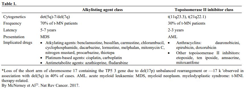

Table 1 Table 1

Nardi et al.,[5] studying three groups of MDS patients, primitive, radiotherapy related (XRT), and chemotherapy-related (C/CMT), showed that the distribution of MDS types according to the WHO Classification for non–therapy-related (primary) MDS and the blood counts at presentation were similar among the three groups. Only 27% of the XRT patients had intermediate-2 or high IPSS scores, compared with 60% of the C/CMT patients. Patients in the XRT group had IPSS scores significantly lower than the C/CMT patients p<.001) but similar to the de novo (p-MDS) MDS/CMML patients. These authors conclude that patients with t-MN after XRT alone had a superior overall survival (p < .006) and a lower incidence of high-risk karyotypes (p <.01 for AML and < .001 for MDS) compared with patients in the C/CMT group. (Table 2)

Table 2 
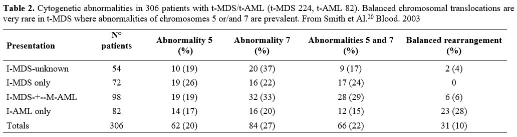
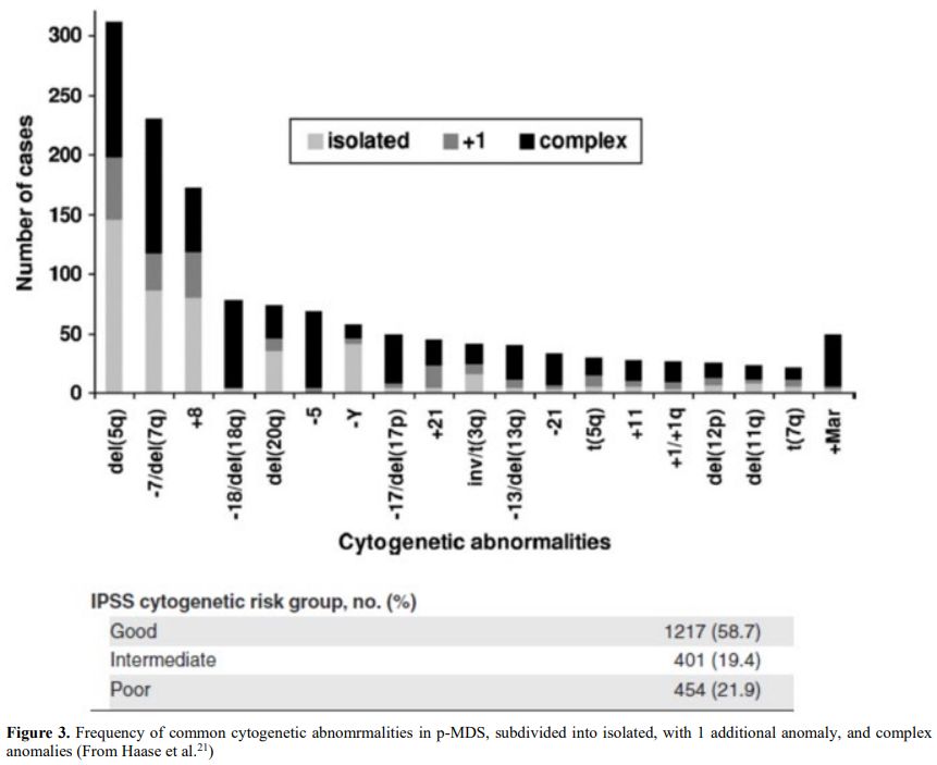
Table 2. Cytogenetic abnormalities in 306 patients with t-MDS/t-AML (t-MDS 224, t-AML 82). Balanced chromosomal translocations are very rare in t-MDS where abnormalities of chromosomes 5 or/and 7 are prevalent. From Smith et Al.[20] Blood. 2003
Table 2.2

In contrast, there were no significant differences in survival or frequency of high-risk karyotypes between the XRT and de novo groups. AML and MDS diagnosed in the past decade in patients after receiving XRT alone differ from t-MN occurring after C/CMT and share genetic features and clinical behavior with de novo AML/MDS. These results suggest that post-XRT MDS/AML may not represent a direct consequence of radiation toxicity and warrant a therapeutic approach similar to de novo disease. Similarly, the MDS secondary to benzene (Bz) also seems to have characteristics not similar or identical to t-MDS/AML, as supposed in the past.[11,12]
Irons et al.[12] recently studied the prevalence of hematopoietic and lymphoid diseases for 2,923 consecutive patients prospectively diagnosed in their laboratory in Shanghai utilizing World Health Organization (WHO) criteria. The Shanghai series of 722 cases of AML includes the most extensive published series with documented Bz exposure and 644 unexposed de novo-AML cases. They also reported the clinical, phenotypic, and molecular characteristics of MDS developing in 649 patients, of whom 80 were determined to have some Bz exposure. In as much as t-MDS and t-AML are considered to be overlapping entities, they initially were surprised to discover that MDS presenting in individuals with chronic exposure to Bz at high concentrations (67–100 mg/m3) did not exhibit a pattern of cytogenetic and phenotypic abnormalities typically observed in t-MDS. In fact, in their MDS series, cases associated with exposure to the highest concentrations of Bz (n 5 29) were found to have a lower prevalence of clonal cytogenetic abnormalities (24%) when compared with unexposed, that is, p-MDS cases (30%) (n 5 569). Further, they did not observe an increase in clonal deletions involving all or part of chromosomes 5 or 7 in Bz-exposed MDS relative to unexposed MDS cases and in no instance involving 5/5q- as the sole abnormality. The conclusion was that benzene exposed MDS-Leukemia patients more closely resemble de-novo than therapy-related MDS leukemia.
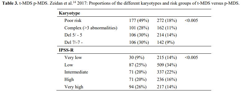
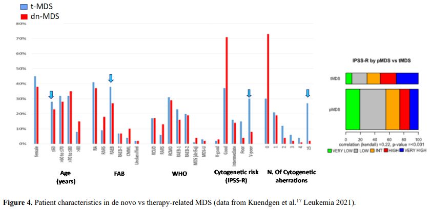
It is also very significant to notice that the prognosis of both MDS de novo and therapy-related can be evaluated by the same scoring systems.[8,14-17] Of the different methods of stratifying t-MDS, the best seems to be the IPSS-R (International Prognostic Scoring System, Revised), which considers, in descending order, five major variables for evaluating clinical outcomes, including cytogenetic risk groups, marrow blast percentage, and depth of cytopenias (hemoglobin, platelet, and ANC levels, respectively), therefore, to affirm that all t-MDS patients have an unfavorable prognosis is not valid. In general, in the t-MDS series, the proportion of high-risk patients is higher.[8,14-17] Furthermore, in general, the survival of every category was lower for t-MDS; however, patients with IPSS and MPSS had a very low survival, not significantly different in both patients with t-MDS and with p-MDS.[15] In the study of Zeidan, patients with t-MDS had significantly inferior survival than p-MDS (median, 19 vs. 46 months, respectively, P=0.005), and patients with t-MDS had a higher prevalence of adverse prognostic factors such as poor risk cytogenetics and higher blast percentages. Although a less favorable clinical outcome occurred in each t-MDS subset compared with p-MDS subgroups, FAB and WHO-classification, IPSS-R, and WPSS-R (WHO-based Prognostic Scoring System-revised) effectively separated t-MDS patients into different risk groups, indicating that all established risk factors for p-MDS maintained relevance in t-MDS, with cytogenetic features having enhanced predictive power. Thus there is significant heterogeneity in clinical outcomes in t-MDS with a small subset of patients having a more indolent disease course, who might not necessarily benefit from aggressive therapeutic interventions.[15,16] All this was confirmed in a large and multicenter study[17] where the predictive power of IPSS-R was almost comparable top-MDS in patients with a solid tumor as primary disease as well as in patients after radiotherapy only. However, the predictive power was lower in patients with a history of hematologic disease treated with chemotherapy. It is important to highlight that this extensive work demonstrated an unexpectedly high percentage of good-risk and normal cytogenetics, concordantly with other more recently published data[8,15,16] Figure 1, 2.


Figure 1 
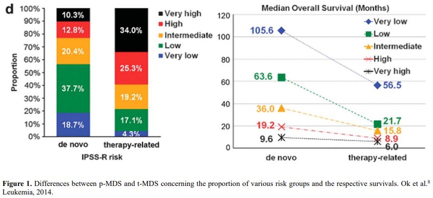
Figure 1. Differences between p-MDS and t-MDS concerning the proportion of various risk groups and the respective survivals. Ok et al.[8] Leukemia, 2014.

Figure 2 
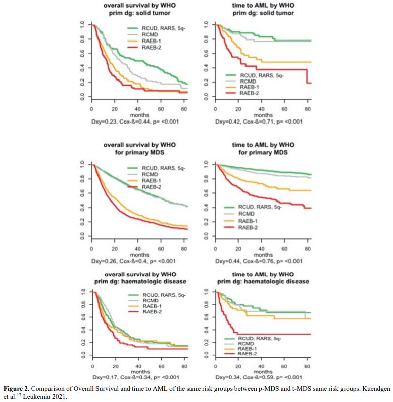
Figure 2. Comparison of Overall Survival and time to AML of the same risk groups between p-MDS and t-MDS same risk groups. Kuendgen et al.[17] Leukemia 2021.


Cytogenetics

Cytogenetics has a fundamental role in determining the prognosis of de novo MDS. However, its importance in t-MDS has been taken into consideration only recently.
Recurrent chromosomal abnormalities are present in 40%–70% of patients with de novo MDS at diagnosis (Figure 3). However, they are present in 90-95% of patients with t-MDS, frequently in the context of complex karyotypes. Frequent chromosomal abnormalities in patients with t-MDS post-alkylating agents include −5/del(5q), −7/del(7q), and/or +8, whereas translocations involving 11q23 or 21q22, as well as t(15;17); Inv 16; t(17;19)(q22;12), have been frequently reported in patients with prior exposure to topoisomerase II inhibitors[18-23] (Table 2, Table 3).

Figure 3 Figure 3. Frequency of common cytogenetic abnomrmalities in p-MDS, subdivided into isolated, with 1 additional anomaly, and complex anomalies (From Haase et al.[21])

Table 3 Table 3. t-MDS p-MDS. Zeidan et al.[14] 2017: Proportions of the different karyotypes and risk groups of t-MDS versus p-MDS.

In t-MDS and p-MDS, the same cytogenetics abnormalities are present. Are they indeed similar, and do they have similar significance and prognosis? Some chromosomal abnormalities are more frequent in t-MDS than in p-MDS; however, there are no specific t-MDS karyotypes (Figure 3, 4). Most chromosomal abnormalities present in t-MDS have in the past been considered to have a poor prognosis, but this is not always true (Table 3).

Figure 4 Figure 4. Patient characteristics in de novo vs therapy-related MDS (data from Kuendgen et al.[17] Leukemia 2021).


Abnormalities of chromosome 5

A typical example is a deletion of chromosome 5, which is considered favorable in p-MDS but unfavorable in t-MDS.[24-26] To justify this discrepancy, Le Beau showed among others that there are two minimally deleted regions (MDRs) on chromosome 5q: 5q31.2 in patients with t-MN, de novo AML, and high-risk MDS; and 5q32 in those with 5q– syndrome, now classified as MDS with isolated del(5q), which has a good prognosis.[22,24] It is essential to distinguish the simple deletion of chromosome 5 from the syndrome of 5q-;[24] furthermore, it should be mentioned that the deletion of chromosome 5 is frequently associated with another chromosome abnormality in the MDS therapy-related, as -7/del 7, and this worsens the prognosis.[22,24] However, some authoritative papers report no differences between isolate del(5q) in t-MDS and p-MDS. Lessard et Al.[27] report that the breakpoints for 5q vary,18 mainly from 5q11 to 5q35, so the deletion size may be small (mainly 5q31), medium (size equivalent to half of the long arm, whatever bands are involved), or large (almost all the long arm). Thus, they distinguished 19 cases with small (17%), 27 cases with medium (25%), and 64 cases with large (58%) deleted segments. They found no difference in the distribution of these deletions in 28 therapy-related cases, with 6 small (21%), 5 medium (18%), and 17 large deleted segments (61%). Similarly, Holtan and Al.,[28] in their series of 130 patients with 5q deletion, of which 32 were therapy-related, found that the breakpoints defined by G-banded karyotyping poorly correlate with particular disease features. Surprisingly, the survival of patients with treatment-related MDS was equivalent to those with de novo MDS and del(5q). Morphologic features associated with del(5q) are diverse. Most patients with del(5q) MDS do not meet the criteria for WHO-defined 5q-syndrome, and the presence of del(5q) does not appear to modify the clinical phenotype. Consequently, in the IPSS-R applied to t-MDS, as for p-MDS, the 5q- appears to be a favorable factor.[15-17] However, the effects of 5 deletion is influenced by additional mutations, including SF3B1and TP53.[29]

Chromosome 7 abnormalities

Isolated monosomy 7 (−7), and/or partial loss of the long arm of chromosome 7 (del 7q) and –7/del(7q) with additional cytogenetic aberration(s) are the second most frequent chromosomal abnormalities in p-MDS and are associated with poor OS and high AML transformation rate. In t-MDS, it plays a similar unfavorable prognostic role; they represent the most frequent cytogenetic abnormalities and are frequently associated with additional cytogenetic aberration, as 5q- deletions.[17,19,20]
Recently Crisà[30] et al. reported a large series of myelodysplastic syndrome patients collected from some European centers with partial or total loss of chromosome 7. Of this series (280 patients), 32 had a t-MDS, and the outcome did not differ from that with p-MDS. The median number of mutations per patient was 2 (range 0–8). Patients harboring ≥2 mutations had a worse outcome than patients with <2 or no mutations (leukemic transformation at 24 months, 38%, and 20%, respectively, p=0.044). Untreated patients with del(7q) had better overall survival (OS) compared with those with −7 (median OS, 34 vs. 17 months, p=0.034). In multivariable analysis, blast count, TP53 mutations, and number of mutations were independent predictors of OS, whereas the cytogenetic subgroups did not retain prognostic relevance.[30]
However, del(7q) detection in patients following cytotoxic therapies is not always related to an emerging therapy-related myeloid neoplasm. Goswami et Al.[31] described 39 patients who acquired del(7q) as sole abnormality following cytotoxic therapies for malignant neoplasms. The median interval from cytotoxic therapies to del(7q) detection was 40 months (range, 4-190 months). Twenty-eight patients showed an interstitial, and 11 a terminal 7q deletion. Fifteen patients (38%) had del(7q) as a large clone and 24 (62%) as a small clone. With a median follow-up of 21 months (range, 1-135 months), 18 (46%) patients developed a therapy-related myeloid neoplasm, including all 15 patients with a large del(7q) clone and 3/24 (12.5%) with a small clone. Of the remaining 21 patients with a small del(7q) clone, 16 showed no evidence of t-MN, and 5 had an inconclusive pathological diagnosis. The authors conclude that isolated del(7q) emerging in patients after cytotoxic therapy may not be associated with t-MN in about half of patients. The clone size of del(7q) is critical; a large clone is almost always associated with therapy-related myeloid neoplasms, whereas a small clone can be a clinically indolent or transient finding.[31]

Chromosome 17 abnormalities

Chromosome 17 (chr17) abnormalities are found in about 2% of patients with de novo myelodysplastic syndrome and about 4.5 % of t-MN.[32,33] In a Spanish study,[32] chromosome 17 abnormalities were classified into three groups: isolated 17 (20.5%), with one additional abnormality (8; 9.1%), and two or more additional abnormalities (complex karyotype) (70.4%). Overall survival and transformation to AML were the same in the first two groups, while there was a significant difference with the patients with complex karyotype.
The Fenaux group,[33] between 1982 and 1996, performed a cytogenetic analysis in 755 cases of MDS and 754 cases of AML, classified according to FAB criteria. Sixty-nine of them (4.6%) had cytogenetic rearrangements leading to 17p deletion, with 25 (36%) occurring after a primary neoplasm treated with chemotherapy and/or radiotherapy; 21 were classified, according to FAB, as MDS, and only 4 as AML. Eighteen patients had unbalanced translocation between chromosome 17 and another chromosome, including chromosome 5 in 8 cases, 7 in three cases, and 1 in one.
Most frequent primary neoplasms were hematologic, with a high prevalence of Philadelphia-negative myeloproliferative neoplasms-, treated with alkylating agents, hydroxyurea or radioactive phosphorus:[33] About half of the patients, in addition, were typical in that they occurred mainly after lymphoid malignancies treated with alkylating agents, and also had monosomy 7 or del 7q. However, the remaining half unexpectedly occurred in ET or PV treated mainly by hydroxyurea, 32P, or pipobroman and rarely presented chromosome 7 abnormalities. p53 mutations and/or abnormal p53 expression were found in 16 of the 19 evaluable cases.
Isolated isochromosome (17q) has also been reported in patients with primary or therapy-related MDS/MPN and AML by the MD Anderson Cancer Center.[34] Myeloid neoplasms with isolated isochromosome 17q represent a distinct clinicopathologic entity with myelodysplastic and myeloproliferative features, high risk of leukemic transformation, and wild-type TP53.[34] The Hellenic 5‐azacytidine registry[35] of 548 adult patients with MDS treated with 5‐azacytidine reports 32 patients with a chromosome 17 abnormality (6 with i[17q], 15 with ‐17, 3 with add[17p] and the rest with other rarer abnormalities, mostly translocations). The presence of a chromosome 17 abnormality was associated with poor prognostic features (high IPSS, IPSS‐R, and WPSS scores) and a low overall survival rate (15.7 vs. 36.4 months for patients without chromosome 17 abnormalities. The Brit et Al.[36] study reported the long-term outcomes of 98 patients with AML or MDS with chromosome 17 abnormalities, 55 had de novo MDS/AML, and 43 had secondary MDS/AML. There were no differences between the two groups regarding the presence of monosomal karyotype (69.1 % versus 67.4 %; p = 0.51). Similarly, there was no significant difference in the prevalence of complex karyotype (98.1 % versus 95.3 %; p = 0.41), or of ch17 abnormalities at diagnosis between the two groups (87.3 % versus 83.7 %; p = 0.41).

Chromosome 20

20q deletion is common in MDS, represents about 5% of all cases of p-MDS, and is considered associated with a good prognosis. Braun et al.[37] have reported for the Groupe Francophone des Myélodysplasies (GFM) over almost 20 years, 62 cases of isolated chromosome 20 deletion and 36 patients with del 20q and other cytogenetic abnormalities, and 1335 MDS patients without del20q. Characteristics of these patients were: lower platelet, a low proportion of marrow blast, and high reticulocyte counts. Median survival was 54 months in patients with isolated del 20q, not reached, and 12 months for del 20q with one or several additional abnormalities, respectively (p = 0.035), confirming the favorable prognosis of del20q without complex abnormalities. Kanagal-Shamanna et al.[38] from MD Anderson Cancer Center identified five cases of t-MN and 26 cases of de novo MDS with isolated del(20q) over ten years.[38] All cases had a long latency interval from the treatment of the primary malignancy to the onset of t-MN, and all were associated with frequent bone marrow dysplasia. del(20q) was the sole abnormality detected at the time of diagnosis of t-MN in three cases, six years before diagnosis in one case, and at the time of relapse of AML in one case. Three patients with t-MDS had a relatively indolent clinical course, whereas two presented with AML or developed AML shortly after t-MDS. The patients with de novo MDS and isolated del(20q) frequently presented with anemia and thrombocytopenia associated with bone marrow dysplasia. The median overall survival was 64 months.[38] Frequently, these patients had minimal signs of dysplasia and an indolent course.[39]
Yin, Peng, Shamanna et Al.[40] identified 92 patients who acquired isolated del(20q) following cytotoxic therapies for malignant neoplasms. Seventy-six patients showed interstitial, and sixteen patients showed terminal 20q deletion. The median interval from prior cytotoxic therapies to del(20q) detection was 58 months (range, 5-213 months). With a median follow-up of 23 months (range, 1-183 months), 21 (23%) patients developed a t-MN, and 71 (77%) patients did not. In patients who developed a t-MN, del(20q) was present in a higher percentage of metaphases (60 vs. 25%, P<0.0001); persisted for a longer period (24 vs. 10 months, P=0.0487); and was more often a terminal deletion (33 vs. 13%, P=0.0006) compared with patients who did not develop t-MN. Clonal evolution was only detected in t-MN (4 patients, 19%).
These data show that del(20q) emerging after cytotoxic therapy represents an innocuous finding in more than two-thirds of patients. However, in patients who develop a t-MN, del(20q) often involves a higher percentage of metaphases, persists longer, and more frequently is a terminal rather than an interstitial deletion.
Cameron Yin et al.[41] reported 64 patients with chronic lymphocytic leukemia (CLL) and del(20q) as the sole abnormality in 40, a stemline abnormality in 21, and a secondary abnormality in 3 cases. Fluorescence in situ hybridization (FISH) analysis revealed an additional high-risk abnormality, del(11q) or del(17p), in 25/64 (39%) cases. In most cases, the leukemic cells showed atypical cytologic features, unmutated IGHV (immunoglobulin heavy-chain variable region) genes, and ZAP70 positivity. The del(20q) was detected only after chemotherapy in all 27 cases with initial karyotype information available. With a median follow-up of 90 months, 30 patients (47%) died, most due to CLL. Eight patients developed a t-MN, seven with a complex karyotype. In 12 cases without morphologic evidence of a myeloid neoplasm, combining morphologic and FISH analysis, localized the del(20q) to the CLL in 5 (42%) cases and to myeloid/erythroid cells in 7 (58%) cases. The del(20q) was detected in myeloid cells in all 4 cases of MDS. Together, these data indicate that CLL with del(20q) acquired after therapy is heterogeneous. In the cases with morphologic evidence of dysplasia, the del(20q) likely resides in the myeloid lineage. However, in cases without morphologic evidence of dysplasia, the del(20q) may represent clonal evolution and disease progression. Combining morphologic analysis with FISH for del(20q) or performing FISH on immunomagnetically selected sub-populations to localize the cell population with this abnormality may help guide patient management.
Nilsson et al.,[42] analyzed chromosome abnormalities in MM/MGUS, including 122 MM and 26 MGUS/smoldering MM (SMM) cases. Sixty-six (54%) MMs and 8 (31%) MGUS/SMMs were karyotypically abnormal. Of these, 6 (9%) MMs and 3 (38%) MGUS/SMMs displayed myeloid abnormalities, that is, +8 (1 case) and 20q- (8 cases) as the only anomalies, without any evidence of MDS/AML. One patient developed AML, whereas no MDS/AML occurred in the remaining eight patients. In one MGUS, FISH analyses revealed del(20q) in CD34+CD38- (hematopoietic stem cells), CD34+CD38+ (progenitors), CD19+ (B cells), and CD15+ (myeloid cells). These data indicate that 20q- occurs in 10% of karyotypically abnormal MM/MGUS cases and might arise at a multipotent progenitor/stem cell level. Patients with sole del(20q) chromosomal abnormality and without morphologic features of a MN display variable clinical outcomes. To explore the potential risk stratification markers in this group of patients, Ravindran et Al. evaluated the mutational landscape by a 35-gene MN-focused next-generation sequencing (NGS) panel and examined the association of mutations to MN progression.[43] Fifty-five patients with isolated del(20q) were studied over ten years, of whom 23 (41.1%) harbored at least one mutation, and 16.1% of them progressed into MNs during follow-up. Thus, all patients who progressed harbored one or more pathogenic mutations at the time of isolated del(20q) diagnosis, and the presence of mutations was a statistically significant risk factor for MN progression. Additionally, among the 23 patients harboring mutations, DTA epigenetic modifier mutations (DTA: DNMT3A, TET2, and ASXL1) did not influence progression, whereas mutations occurring in the non-DTA genes were differentially distributed in patients with and without progression. The mutations involving non-DTA genes include IDH1, IDH2, and BCOR, kinases CBL, PTPN11, JAK2, tumor suppressors TP53 and PHF6, and transcription factor RUNX1.
In conclusion, several studies show that chromosome 20 deletion in both de novo and t-MDS has a favorable prognostic significance when isolated, and its presence does not necessarily bring about a t-MN.

Somatic Mutations

Since traditional morphology, cytogenetics, and flow cytometry failed to identify clinical features differentiating t-MDS from p-MDS, several authors are trying to define a molecular landscape able to distinguish these two subgroups of MDS according to the presence of somatic mutations in genes known to be involved in myeloid neoplasm pathogenesis. Accordingly, 78 to 90% of MDS patients have one or more oncogenic mutations. Furthermore, de novo MDS show a mutational profile prevalently characterized by a high frequency of somatic mutations in genes involved in spliceosome machinery[44,45] (prevalently, SF3B1, SRSF2, U2AF1, and ZRSR2), DNA methylation regulators (DNMT3A, TET2, IDH1and IDH2), histone modifiers (ASXL1 and EZH2), transcription factors (RUNX1, TP53, and others) and signal transduction genes (JAK2, KRAS, CBL).[45,46]
Mutations in the spliceosome machinery gene SF3B1 (Splicing Factor 3b Subunit 1) were closely associated with specific MDS subgroups with ring sideroblast (RS).[45.47] The recently published data extrapolated from the dataset of the IWG-PM (International Working Group for the Prognosis of MDS) strongly confirm the enrichment of these mutations in refractory anemia with RS (RARS) and refractory cytopenia multilineage dysplasia with RS (RCMD-RS) in 82% and 75% of cases, respectively.[48] Due to the tight association of SF3B1 mutations with the disease phenotype of RS,[49,50] according to “The 2016 revision to the World Health Organization classification of myeloid neoplasms and acute leukemia”, a diagnosis of MDS-RS can be made if ring sideroblasts are >5% of nucleated red cells, and a somatic mutation of SF3B1 is present, instead of 15% of RS without mutation.[1]
Several reports suggest that SF3B1 mutations have a good prognostic value on overall survival (OS) and risk of disease progression, and these findings were recently confirmed in very low and low-risk IPSS-R categories by the analysis of IWG data set.[46-50]
Although t-MN and p-MDS patients with very low or low IPSS-R show similar biological and clinical characteristics, the RS phenotype in t-MN is not associated with improved survival, even if mutations in spliceosome machinery genes have been identified at higher frequency in p-MDS compared to t-MN (56.5% vs. 25.6%, respectively)51 and among genes belonging to this pathway, SF3B1 and U2AF1 are more frequently mutated in p-MDS (25.9% vs. 6.2% and 7.4% vs. 1.6%, respectively). In contrast, mutations in SRSF2 and ZRSR2 genes seem equally distributed. Singhal et al. showed a similar frequency of RS in their p-MDS and t-MN cohorts (29.6% vs. 24.8%), but RSs were 15% of erythroid cells in 21.3% of p-MDS and in only 14.7% of t-MN.[51] Remarkably, mutations in spliceosome pathway were identified in all patients with p-MDS and 15% of RS (SF3B1, 96%), but only in 37% of t-MN with 15% of RS (SF3B1, 32%), showing a less significant association of these mutations with the RS phenotype. Moreover, the authors identified TP53 mutations in 29.5% of the entire cohort of t-MN patients and, unexpectedly, in 92% of those with 15% RS and no SF3B1 mutations, highlighting a new potential role of TP53 mutations in the context of t-MN with RS. In this line, the median OS of t-MN with 15% RS (12 months) was significantly lower than that of p-MDS with 15% RS (38 months), pointing out the poor prognostic role of TP53 mutations also in t-MN patients with RS and the weak association with SF3B1 mutations.[51] The group of MD Anderson Cancer Center reported similar data:[52] Commonly mutated genes were TP53 (56.5%), TET2 (39.1%), SF3B1 (35.7%), ASXL1 (30.4%), DNMT3A (17.4%), RUNX1 (17.4%) and SRSF2 (14.3%). Compared with d-MDS-RS, TP53 mutation was more common, but SF3B1 mutation was less common in t-MDS-RS (p < 0.05). In t-MDS-RS, Mutations in 4 genes (SF3B1, U2AF1, SRSF2, and ZRSR2) involving RNA splicing were found in about 50% of patients compared to ˜90% in d-MDS-RS. Overall survival was by far worse in t-MDS-RS compared to d-MDS-RS (median overall survival: 10.9 months and 111.9 months in t-MDS-RS and p-MDS-RS, respectively, p < 0.05). Progression to AML was more common in t-MDS-RS (18.4% vs. 7.4% in t-MDS-RS and d-MDS-RS, respectively, p < 0.05). Unlike de novo MDS, t-MDS-RS did not have a different outcome than t-MDS without RS (median OS: 10.9 months vs. 14.3 months, respectively, p=0.2341). Mutation profiles suggest that RS in t-MDS might be a secondary event in at least 50% of the cases or not related to mutations in RNA splicing machinery, unlike p-MDS, where they occur early and are associated with ineffective erythropoiesis. Although in the last decade, the mutational profiles of p-MDS and t-MN have been extensively analyzed, identifying 79% to 95% of patients mutated in at least one gene, without significant differences in cohorts, and none of the studied genes exclusively mutated in one of them, the prevalence of cases mutated in specific genes seem to be different.[52,53,54]
Today, mutations in the TP53 gene are considered the most frequent molecular alterations identified in t-MDS/t-MN patients, with a frequency from 30 to 47%, according to both specific characteristics of the study cohort and depth of sequencing technologies.[51-56] In these series of patients, authors also identified a tight association of TP53 mutations with the presence of complex karyotype, defined as three chromosomal abnormalities, in about 80% of cases, and a poor prognosis compared to TP53 wild-type t-MN patients. In 2017, Lindsley et al., screening through t-NGS technology the mutational profile of a large cohort of 1514 MDS patients undergoing Hematopoietic Stem Cell Transplantation (HSCT), found interesting differences in the frequency of TP53 mutations stratifying patients according to p-MDS and t-MDS (311 patients) subgroups.[53] They found a higher prevalence of TP53 mutations in t-MDS than p-MDS (38% vs. 14%, respectively) and significantly shorter survival. Of note, in t-MDS, the authors also identified a higher frequency of mutations in the TP53 regulator PPM1D compared to p-MDS (15% vs. 3%, respectively), reaching the quote of 46% of mutations in TP53 or PPM1D.
Furthermore, the co-occurrence of TP53 and PPM1D mutations was higher in t-MDS than stochastically expected in patients with PPM1D mutations alone (51%), when compared with PPM1D and TP53 double wild-type patients, had a similar frequency as those with TP53 mutations alone (39%) or double mutated PPM1D and TP53 (54%).[53] Although PPM1D gene encodes a serine-threonine protein phosphatase, involved in the cellular response to environmental stress and therefore to exposure to leukemogenic therapies, through the inhibition of TP53 activity, PPM1D mutated t-MDS patients without TP53 mutations did not show any association with the presence of complex karyotypes and adverse prognosis. Even if more frequent than p-MDS, TP53 mutation characteristics in t-MDS and t-AML are similar to de novo diseases.[55] Consistent with the tumor-suppressive role of TP53, patients may harbor both mono- and biallelic mutations. The International Working Group for Prognosis in MDS[56] assembled a cohort of 3,324 with MDS and studied the effect of TP53 allelic state on genome stability, clinical presentation, outcome, and response to therapy.56 Outcomes of monoallelic cases significantly differed with the number of co-occurring driver mutations; for example, the 5-year survival rate of monoallelic patients with no other identifiable mutations was 81%, while it was 36% for patients with one or two other mutations, 26% for patients with three or four other mutations, and 8% for patients with more than five other mutations.
Other than prognostic predictors of outcome, acquired somatic mutations may also play a pivotal role in the context of MDS susceptibility. In 2014 a real earthquake shocked the world of hematologists redefining the borders between hematology and onco-hematology by discovering acquired somatic mutations in the same genes previously known to be involved in myeloid neoplasms in older people without traces of hematologic disorders. In independent works, screening by whole-exome sequencing (WES) in large cohorts of non-hematological patients, Genovese and Jaiswal, identified a close association between hematopoietic stem cell aging and the accumulation of somatic mutations in patients without hematological malignancies.[57,58] These mutations, now known as DTA (DNMT3A, TET2, and ASXL1), have been found enriched in a linear relationship with age, with about 20% of mutations in people 80 years old. Although DTA genes were the most enriched, other genes such as JAK2, SF3B1, PPM1D, and TP53 were found enriched in a linear relationship with age, while others such as FLT3, NPM1, IDH1, and IDH2 were rarely found mutated in these subjects.[59] These scenarios of clonal hematopoiesis, now defined as ARCH (Age-related clonal hematopoiesis) or CHIP (Clonal hematopoiesis of indeterminate potential) according to the VAF of identified mutations (without a specific cut-off or 2%, respectively),[59] have been linked to an overall increased risk of transformation to hematological malignancy, with a risk of progression of about 0.5–1% per year vs. <0.1% in non-CHIP carriers, and may represent a pre-malignant state in p-MDS and t-MN, whose development can be triggered by exposure to cytotoxic damage.[57,58,60] In this line, several authors were able to identify pre-existing somatic mutations, at very low VAF, in patients who developed a t-MN after a chemo/radiotherapeutic treatment for a primary tumor or an autoimmune disease, demonstrating the expansion of CHIP mutations and their role as a predisposing factor for MN development.[61-66] Very recently, Zeventer et al., using high-throughput t-NGS technology (gene panel of 27 driver genes at a VAF of 1%), redefined the prevalence of clonal hematopoiesis (CH) in a cohort of 621 individuals aged 80 years extrapolated from the LifeLines cohort (Netherlands database of 167,729 participants).[67] Clonal hematopoiesis was identified in the peripheral blood of 61.5% of individuals, without differences across gender. DNMT3A was mutated in 219 of 621 individuals (35%), 27% with multiple mutations. Similar results were reported for TET2 (166 of 621 individuals (27%), with multiple mutations in 24% of cases, while ASXL1, spliceosome machinery, and TP53 variants were identified at lower frequencies (6%, 4%, and 3%, respectively). Although CH was not associated with a higher risk of death in the complex, individuals carriers of mutations in genes other than DNMT3A and TET2 were at higher risk than wild-type controls (HR, 1.48; CI, 1.06-2.08). In the same line, the authors did not identify any association between elevated risk of exposure to DNA damaging toxicities (job-related exposure to pesticides, exposure to cytotoxic and/or radiation therapy for primary cancer, and smoking status), and both the prevalence of CH and the number of somatic variants.

Figure 5 Figure 5.  Mutational profiles in myeloid neoplasms. Mutational profile of therapy-related myelodysplastic syndromes (t-MDS) versus de novo MDS. Asterisk denotes genes with a significant difference between t-MDS versus p-MDS.  Ok et al. [56] Leukemia Res., 2015.

In contrast, mutations in selected driver genes, such as spliceosome machinery and ASXL1, were identified at higher frequencies in exposed individuals (6% vs. 1% and 7% vs. 2%, respectively).[67] In this line, our group recently showed that patients CLL who developed a t-MN following treatment with chemoimmunotherapy presented prior to treatment start mutations in CHIP-related genes at a significantly higher prevalence than a large cohort of control CLL. These data show that CHIP increases susceptibility to t-MN also in the CLL context.
In the last few years, a rare mutation in MDS, the Nucleophosmin (NPM1) mutation common in AML and associated with high remission rates and prolonged survival with intensive chemotherapy, has assumed a particular significance.[69] NPM1 mutations are rare in syndromes p-MDS (MDS/MPN), representing about 2% of all MDS.[70,71] The different outcomes of these patients, if treated with intensive chemotherapy or demethylating agent, induce to combine them in a subgroup. The NPM1 mutations have been found even more rarely in t-MDS and more frequently in t-AML.[71,72] Andersen et al. observed NPM1 mutations in 7 of 51 patients presenting as overt t-AML, as compared to only 3 of 89 patients presenting as t-MDS (P<0.037); only 1 of 10 patients with NPM1 mutations presented chromosome aberrations characteristic of the therapy-related disease, and 7q-/-7, and 5q-/-5 the most frequent abnormalities of t-MDS/t-AML, were not observed (P<0.002). This raised the question of whether some of the cases presenting NPM1 mutations were, in fact, cases of de novo leukemia or de novo MDS.[72] This same issue is relevant in p-MDS, where the presence of NPM1 mutations is associated with an increased rate of AML transformation, raising the question of whether these MDS indeed represent AML at an early stage.
All these data highlight the potential role of acquired mutations and/or cytogenetic abnormalities in the definition of de novo and therapy-related MDS, which will have to be taken into account in the next editions of the WHO classification.

Epigenetic Regulation

Changes in gene expression not directly linked to changes in DNA sequence constitute the fundamental concept of epigenetic regulation. Aberrant differentiation in MDS can often be traced to an abnormal epigenetic process represented by both gains and losses of DNA methylation genome-wide and at specific loci, as well as mutations in genes that regulate epigenetic programs, such as TET2 and DNMT3a, involved in DNA methylation control, and EZH2 and ASXL1, involved in histone methylation control.[72]
Epigenetic changes include various covalent modifications of nucleic acids and histones, which finely coordinate the gene expression of single cells, defining their role and phenotype. These regulatory mechanisms mainly include DNA methylation, histone modifications, and chromatin remodeling. Alterations in epigenetic regulation have been tightly associated with cancer development and progression.[74] Moreover, since all these mechanisms are reversible, the involved genes are considered important epigenetic targets for drug development and patient treatment. Of note, many genes involved in epigenetic regulation, such as DNA methylation regulators (DNMT3A, TET2, IDH1, and IDH2) and histone modifiers (ASXL1 and EZH2), have been identified as being frequently mutated in MDS.[44-46]
Epigenetic regulation through DNA methylation is mainly driven by DNA methyltransferase enzymes (DNMTs) by the addition of a methyl group (CH3) to a cytosine residue (5-methylcytosine) within a CpG dinucleotide.[75] Regions enriched in CpG dinucleotides (CpG islands) have been found in the promoter region of about 50% of human genes, but a methylation enrichment was also found in enhancers and transcription factor binding sites.[76]
The nucleoside analog azacytidine (AZA, Vidaza®, Celgene) and its deoxy derivative decitabine (DAC, Dacogen®, Janssen)are able to stimulate gene expression through DNA hypomethylation, following irreversible DNMTs sequestration.[77] However, this action is reversible since inhibitors do not influence de novo DNMTs synthesis. In this line, de novo DNMTs synthesis could explain the rapid relapse after treatment interruption and the requirement for maintenance treatment as long as the response persists.[78,79]
Several authors tried to identify specific DNA sequences or methylation profiles useful as a marker of response to hypomethylating agents that may help to support the decision to continue or stop hypomethylating treatment.
Although gene-specific hypermethylation has been identified as a negative prognostic factor in hematological malignancies, there is no complete agreement on the role of specific candidate genes.
Among these, genes belonging to lineage commitment, apoptosis, cell cycle, immune response, signal transduction, and cytoskeletal remodeling are hypermethylated in MDS patients, and their expression could be modulated by hypomethylating treatment.[80-85]
In addition, the hypermethylation of some promoter genes, like DAPK1, E-cadherin, and thrombospondin-1, has been reported by several groups, including ours, as more frequent in t-MDS than p-MDS.[86]
Recently, Reilly et al. characterized the methylation profile of 114 bone marrow DNA samples from MDS patients using the bisulfite padlock probe (BSPP) sequencing method to highlight differentially methylated regions of genes belonging to different cellular pathways.[87]
Their results showed five unique methylation clusters, resulting from the bioinformatics analysis “OncoGenic Positioning System Onco-GPS”, useful to sub-classify MDS patients according to their methylation profile. In particular, each methylation cluster was enriched for specific genetic variants and cytogenetic alterations, although different mutational profiles may share the same methylation state. Interestingly, their data showed that the variants in splicing factor genes were mutually exclusive and enriched in specific methylation subgroups.[87] These authors did not observe statistically significant associations between specific methylation clusters and clinical laboratory parameters such as cytopenia or bone marrow blasts percentage. On the other hand, the overall survival curves showed differences in patients belonging to specific methylation sub-groups, identifying two major patterns, demonstrating that, although genetic variants alone are insufficient for determining the methylation profile of MDS patients, the latter could be supplemental information for the prognostic stratification.
Finally, they observed enrichment of cluster-specific methylated regions not only in CpG islands belonging to promoter regions but also in distal regulatory regions of the epigenome. Indeed, results showed an equal or greater number of differentially methylated regions outside promoters.
More recently, Cabezón et al. performed a supervised global methylation analysis of 75 high-risk MDS and secondary AML patients included in CETLAM SMD-09 protocol and treated with HMA or intensive treatment according to age, comorbidities, and cytogenetics. They were able to identify a methylation signature defined by 200 probes, useful to distinguish responder from non-responder patients under AZA treatment.[88]
The same authors also identified a methylation pathway that differentiated patients according to their survival. In contrast, the methylation signature detected at the time of diagnosis was not useful for distinguishing patients who would relapse or progress.[88]
In this study, high-risk MDS and sAML showed a more heterogeneous pathway of methylation than age-matched healthy controls, and this heterogeneity precluded the separation of the patients into distinct sub-groups.
While many studies have explored the epigenome of de novo MDS, little is known about the epigenetics of t-MN and, in particular, t-MDS.

Treatment

The therapy for MDS depends on the stage of the disease, age, comorbidities, and infections.[89-91] The stage has been historically risk-stratified using the International Prognostic Scoring System (IPSS) for MDS,[91] updated with the revised-IPSS (IPSS-R), in more recently by the molecular IPSS-M and the Euro MDS scores.[13,92] The disease stage is risk-based on cytogenetic abnormalities, the degree of cytopenias, and the percentage of bone marrow blasts. The IPSS-R subdivides patients into five groups (very low-, low-, intermediate-, high-, very high-risk) that differ in survival and risk of leukemic transformation. This classification is clinically relevant as the treatment approach differs between higher-risk and lower-risk patient subgroups.[89] However, it has been well recognized that some patients with lower-risk (LR)-MDS using IPSS or IPSS-R do not fare well. Mutations are not considered in the risk stratifications of IPSS and are particularly important in worsening the prognosis in patients with normal cytogenetics.[92,93,94] Therefore, inherent limitations of the current prognostic tools should be recognized in making clinical decisions for individual patients. Integrating molecular assessment in risk stratification tools in the Euro-MDS and IPSS-M scores will likely increase their precision and utility in clinical practice.[92-94] Therapy-related MDS have been classified together with the t-AML in the group of t-MN and considered to have the same poor prognosis, independently of the number of blasts found in the marrow.[1,7] However, in the last few years, it appeared more and more evident that the prognosis of the t-MDS is well evaluated utilizing the same parameter employed for p-MDS.[8,14,16] At variance with p-MDS,[21] most patients with t-MDS belong to high or very high risk categories, independently of the origin.[14,16] (Figure 1, 2; Table 3). The treatment goals of p-MDS and t-MDS are the same: to alter the natural history of the disease, decrease the risk of leukemic progression, and improve survival. However, the only cure of either p-MDS and t-MDS remains the allogeneic hematopoietic cell transplant (HSCT), recommended option in such patients if they are candidates for the procedure, following p-MDS established criteria.[95,96] An unanswered question is whether the transplant should be performed after the high-dose chemotherapy or immediately; a third hypothesis could be after the azacytidine bridge.[97] In any case, like all treatments, HSCT is deeply influenced by cytogenetics and mutations.[53,56] In particular, the mutations P53, frequently associated with complex karyotypes, are strong negative prognostic factors in younger patients.[53,56]
Nevertheless, most patients are not candidates to transplant, so patients with p-MDS or t-MDS are most often treated with the hypomethylating agents, azacytidine or decitabine.[98-108] The hypomethylating agents are both approved for MDS therapy by FDA and EMA and are similarly effective in p-MDS and t-MDS in strict relationship with cytogenetics and mutations found in the patient. Klimek et al.[100] report results in 42 patients with therapy-related MDS treated with either 5-azacitidine (AC) or 21-deoxy-5-azacitidine (DAC). Patients were 25–85 (median 70) years of age and covered the entire spectrum of MDS disease stages. As expected, 69% of patients had poor-risk cytogenetics by IPSS criteria.[8,14] The overall response rate was 38%, and complete remissions occurred in six patients (14%). The median overall survival from the start of therapy was 9.2 months. Thus, these results are very similar, if not identical, to what has been reported with AC for all-comers, most of whom have been patients with de novo MDS.
The demonstrated efficacy of demethylating agents has induced researchers to improve their efficacy by adding other drugs; the anti-PD-1 antibodies seem to be the most promising.[108-109] Furthermore, a series of targeted therapies are under investigation for HR-MDS patients, such as the IDH1 and IDH2 inhibitors evaluated in clinical trials with ivosidenib and enasidenib, respectively.[110,11] Furthermore, there is a promising pipeline of novel agents under active investigation in HR-MDS. For patients with LR-MDS, the treatment goals are to improve their quality of life by managing the underlying cytopenias and side effects. The majority of patients are anemic at presentation, representing a major clinical challenge.[3] Treatment options in this group of patients include active surveillance, red blood cell (RBC) transfusions, erythropoiesis-stimulating agents (ESAs), lenalidomide in patients with del (5q), hypomethylating agents also as oral therapy (HMAs), luspatercept in patients with ring sideroblasts, and immunosuppressive agents in a select group of patients.[110,111]
However, there is no experience of these new drugs in t-MDS, which constitutes about 10% of all MDS.[108] In addition, therapy-related MDS patients are often excluded from therapeutic clinical trials, so a comparison between de novo and therapy-related MDS outcomes with the same stage and therapies is difficult to find in the literature.
Borate et al.[113] in their initial search, examined 1148 therapeutic clinical trials on MDS; however, excluding the trial, which did not mention t-MDS (831), and trials that excluded specifically t-MDS, they found only 18 studies (5.7%) that accrued 231 t-MDS patients in total, representing 3.2% of the total accrued MDS trial patient population. In addition, fewer t-MDS patients were accrued in therapeutic trials sponsored by pharmaceutical sponsors vs. nonpharmaceutical sponsors (2.8% vs. 4.0%; P 5 .0073). This pattern of exclusion continues in actively enrolling trials; only 5 (10%) of 49 studies specifically mention the inclusion of t-MDS patients in their eligibility criteria. These results indicate that therapeutic MDS trials seem to exclude t-MDS patients, rendering study results less applicable to this subset of MDS patients, who often have poor outcomes.[17,102,106] Another problem is that in the trials, t-MDS are frequently included together with t-AML as t-MN as reported by the WHO classification of myeloid neoplasm.[1-114]


Conclusions

The characterization of t-MDS and t-AML[8,14,17] has been recently improved with the use of advanced molecular techniques, and although sharing some overlapping features, these diseases exhibit substantial differences in molecular and cytogenetic characteristics and clinical presentation. A future updated classification should consider these issues, and the proposal to separate t-MDS from t-AML should be discussed. In this line, therapy-related MDS could become a subgroup of MDS. Indeed, since there is no specific marker for therapy-related MN, the diagnosis is made on an anamnestic base only, and this does not exclude that some cases of p-MDS may present after cytotoxic therapy and be defined t-MDS but still display genetic characteristics of p-MDS.
The same path has been taken in t-AML with recurrent translocations, characterized by t(15;17), t(8;21), or inv(16), which, following recent guidelines, should be treated according to patients’ fitness, and not to a previous history of cytotoxic treatment, since the outcome of these AMLs in similar in de novo and therapy-related forms.

References   

  1. Arber DA, Orazi A, Hasserjian R, Thiele J, Borowitz MJ, Le Beau MM, Bloomfield CD, Cazzola M, Vardiman JW. The 2016 revision to the World Health Organization classification of myeloid neoplasms and acute leukemia. Blood. 2016 May 19;127(20):2391-405. doi: 10.1182/blood-2016-03-643544. https://doi.org/10.1182/blood-2016-03-643544 PMid:27069254
  2. Gale R.P., Bennett J.M., Hoffman F.O. Who has therapy-related AML? Mediterr J Hematol Infect Dis 2017, 9(1): e2017025, DOI: 10.4084/MJHID.2017.025 https://doi.org/10.4084/mjhid.2017.025 PMid:28293413 PMCid:PMC5333730
  3. Invernizzi R., Quaglia F., Della Porta M.G. Importance of classical morphology in the diagnosis of myelodysplastic syndrome. Mediterr J Hematol Infect Dis 2015, 7(1): e2015035, doi: 10.4084/MJHID.2015.035 https://doi.org/10.4084/mjhid.2015.035 PMid:25960863 PMCid:PMC4418392  
  4. Della Porta M.G., Picone C. Diagnostic utility of flow cytometry in myelodysplastic syndromes. Mediterr J Hematol Infect Dis 2017, 9(1): e2017017, doi: 10.4084/MJHID.2017.017 https://doi.org/10.4084/mjhid.2017.017 PMid:28293405 PMCid:PMC5333741  
  5. Michels SD, McKenna RW, Arthur DC, Brunning RD. Therapy-related acute myeloid leukemia and myelodysplastic syndrome: a clinical and morphologic study of 65 cases. Blood. 1985 Jun;65(6):1364-72.  https://doi.org/10.1182/blood.V65.6.1364.bloodjournal6561364 PMid:3857944
  6. Wilson CS, Traweek ST, Slovak ML, Niland JC, Forman SJ, Brynes RK. Myelodysplastic syndrome occurring after autologous bone marrow transplantation for lymphoma. Morphologic features. Am J Clin Pathol. 1997 Oct;108(4):369-77. doi: 10.1093/ajcp/108.4.369. https://doi.org/10.1093/ajcp/108.4.369 PMid:9322588
  7. Singh ZN, Huo D, Anastasi J, Smith SM, Karrison T, Le Beau MM, Larson RA, Vardiman JW. Therapy-related myelodysplastic syndrome: morphologic subclassification may not be clinically relevant. Am J Clin Pathol. 2007 Feb;127(2):197-20 https://doi.org/10.1309/NQ3PMV4U8YV39JWJ PMid:17210514
  8. Ok CY, Hasserjian RP, Fox PS, Stingo F, Zuo Z, Young KH, Patel K, Medeiros LJ, Garcia-Manero G, Wang SA. Application of the international prognostic scoring system-revised in therapy-related myelodysplastic syndromes and oligoblastic acute myeloid leukemia. Leukemia. 2014 Jan;28(1):185-9. doi:10.1038/leu.2013.191. https://doi.org/10.1038/leu.2013.191 PMid:23787392  
  9. Nardi V, Winkfield KM, Ok CY, Niemierko A, Kluk MJ, Attar EC, Garcia-Manero G,Wang SA, Hasserjian RP. Acute myeloid leukemia and myelodysplastic syndromes after radiation therapy are similar to de novo disease and differ from other therapy-related myeloid neoplasms. J Clin Oncol. 2012 Jul 1;30(19):2340-7. doi: 10.1200/JCO.2011.38.7340.  https://doi.org/10.1200/JCO.2011.38.7340 PMid:22585703 PMCid:PMC4979234  
  10. Zhang L, Lan Q, Guo W, Hubbard AE, Li G, Rappaport SM,McHale CM, Shen M, J iZ, Vermeulen R, Yin S-N, RothmanN, Smith MT. 2011. Chromosome-wide aneuploidy study(CWAS) in workers exposed to an established leukemogen, benzene. Carcinogenesis 32:605-612. https://doi.org/10.1093/carcin/bgq286 PMid:21216845 PMCid:PMC3066415  
  11. Irons RD, Gross SA, Le A, Wang XQ, Chen Y, Ryder J, SchnatterAR. 2010. Integrating WHO 2001-2008 criteria for the diagnosis of Myelodysplastic Syndrome (MDS): A case-case analysis of benzene exposure. Chem Biol Interact 184:30-38. https://doi.org/10.1016/j.cbi.2009.11.016 PMid:19941839  
  12. Irons RD, Chen Y, Wang X, Ryder J, Kerzic PJ. Acute myeloid leukemia following exposure to benzene more closely resembles de novo than therapy related-disease. Genes Chromosomes Cancer. 2013 Oct;52(10):887-94. doi: 10.1002/gcc.22084. Epub 2013 Jul 10. PMID: 23840003. https://doi.org/10.1002/gcc.22084 PMid:23840003  
  13. Greenberg PL, Tuechler H, Schanz J, Sanz G, Garcia-Manero G, Solé F, Bennett JM, Bowen D, Fenaux P, Dreyfus F, Kantarjian H, Kuendgen A, Levis A, Malcovati L, Cazzola M, Cermak J, Fonatsch C, Le Beau MM, Slovak ML, Krieger O, Luebbert M, Maciejewski J, Magalhaes SM, Miyazaki Y, Pfeilstöcker M, Sekeres M, Sperr WR, Stauder R, Tauro S, Valent P, Vallespi T, van de Loosdrecht AA, Germing U, Haase D. Revised international prognostic scoring system for myelodysplastic syndromes. Blood. 2012;120(12):2454-65. doi: 10.1182/blood-2012-03-420489. https://doi.org/10.1182/blood-2012-03-420489 PMid:22740453 PMCid:PMC4425443  
  14. Zeidan AM, Al Ali N, Barnard J, Padron E, Lancet JE, Sekeres MA, Steensma DP, DeZern A, Roboz G, Jabbour E, Garcia-Manero G, List A, Komrokji R. Comparison of clinical outcomes and prognostic utility of risk stratification tools in patients with therapy-related vs de novo myelodysplastic syndromes: a report on behalf of the MDS Clinical Research Consortium. Leukemia. 2017 Jun;31(6):1391-1397. doi: 10.1038/leu.2017.33. Epub 2017 Jan 23. https://doi.org/10.1038/leu.2017.33 PMid:28111463  
  15. Berggren MD, Folkvaljon Y, Engvall M, Sundberg J, Lambe M, Antunovic P, Garelius H, Lorenz F, Nilsson L, Rasmussen B, Lehmann S, Hellström-Lindberg E, Jädersten M, Ejerblad E. Prognostic scoring systems for myelodysplastic syndromes (MDS) in a population-based setting: a report from the Swedish MDS register. Br J Haematol. 2018 Jun;181(5):614-627. doi: 10.1111/bjh.15243. Epub 2018 Apr 29. https://doi.org/10.1111/bjh.15243 PMid:29707769
  16. Quintas-Cardama A, Daver N, Kim H, Dinardo C, Jabbour E, Kadia T, et al. A prognostic model of therapy-related myelodysplastic syndrome for predicting survival and transformation to acute myeloid leukemia. Clin Lymphoma Myeloma Leuk 2014; 14: 401-410 https://doi.org/10.1016/j.clml.2014.03.001 PMid:24875590 PMCid:PMC4167474  
  17. Kuendgen A, Nomdedeu M, Tuechler H, Garcia-Manero G, Komrokji RS, Sekeres MA, Della Porta MG, Cazzola M, DeZern AE, Roboz GJ, Steensma DP, Van de Loosdrecht AA, Schlenk RF, Grau J, Calvo X, Blum S, Pereira A, Valent P, Costa D, Giagounidis A, Xicoy B, Döhner H, Platzbecker U, Pedro C, Lübbert M, Oiartzabal I, Díez-Campelo M, Cedena MT, Machherndl-Spandl S, López-Pavía M, Baldus CD, Martinez-de-Sola M, Stauder R, Merchan B, List A, Ganster C, Schroeder T, Voso MT, Pfeilstöcker M, Sill H, Hildebrandt B, Esteve J, Nomdedeu B, Cobo F, Haas R, Sole F, Germing U, Greenberg PL, Haase D, Sanz G. Therapy-related myelodysplastic syndromes deserve specific diagnostic sub-classification and risk-stratification-an approach to classification of patients with t-MDS. Leukemia. 2021; 35(3): 835-849. doi: 10.1038/s41375-020-0917-7. https://doi.org/10.1038/s41375-020-0917-7 PMid:32595214 PMCid:PMC7932916  
  18. Kawankar N, Vundinti BR. Cytogenetic abnormalities in myelodysplastic syndrome: an overview. Hematology. 2011 May;16(3):131-8. doi:10.1179/102453311X12940641877966. https://doi.org/10.1179/102453311X12940641877966 PMid:21669051  
  19. Pedersen-Bjergaard J, Andersen MK, Andersen MT, Christiansen DH. Genetics of therapy-related myelodysplasia and acute myeloid leukemia. Leukemia. 2008 Feb;22(2):240-8. doi: 10.1038/sj.leu.2405078. https://doi.org/10.1038/sj.leu.2405078 PMid:18200041  
  20. Smith SM, Le Beau MM, Huo D, Karrison T, Sobecks RM, Anastasi J, Vardiman JW, Rowley JD, Larson RA. Clinical-cytogenetic associations in 306 patients with therapy-related myelodysplasia and myeloid leukemia: the University of Chicago series. Blood. 2003 Jul 1;102(1):43-52. doi: 10.1182/blood-2002-11-3343. https://doi.org/10.1182/blood-2002-11-3343 PMid:12623843  
  21. Haase D, Germing U, Schanz J, Pfeilstöcker M, Nösslinger T, Hildebrandt B, Kundgen A, Lübbert M, Kunzmann R, Giagounidis AA, Aul C, Trümper L, Krieger O, Stauder R, Müller TH, Wimazal F, Valent P, Fonatsch C, Steidl C. New insights into the prognostic impact of the karyotype in MDS and correlation with subtypes: evidence from a core dataset of 2124 patients. Blood. 2007 Dec 15;110(13):4385-95. doi: 10.1182/blood-2007-03-082404. Epub 2007 Aug 28. https://doi.org/10.1182/blood-2007-03-082404 PMid:17726160
  22. Stoddart A, McNerney ME, Bartom E, Bergerson R, Young DJ, Qian Z, Wang J, Fernald AA, Davis EM, Larson RA, White KP, Le Beau MM. Genetic pathways leading to therapy-related myeloid neoplasms. Mediterr J Hematol Infect Dis. 2011; 3(1): e2011019. doi: 10.4084/MJHID.2011.019. https://doi.org/10.4084/mjhid.2011.019 PMid:21713073 PMCid:PMC3113274  
  23. McNerney ME, Godley LA, Le Beau MM. Therapy-related myeloid neoplasms: when genetics and environment collide. Nat Rev Cancer. 2017 Aug 24;17(9):513-527. doi: 10.1038/nrc.2017.60 https://doi.org/10.1038/nrc.2017.60 PMid:28835720 PMCid:PMC5946699  
  24. Le Beau, M M Cytogenetic and molecular delineation of the smallest commonly deleted region of chromosome 5 in malignant myeloid diseases. Proc. Natl Acad. Sci. USA 9 1993:90; 5484-5488. https://doi.org/10.1073/pnas.90.12.5484 PMid:8516290 PMCid:PMC46745  
  25. Boultwood, J. Fidler C, Strickson AJ, Watkins F, Gama S, Kearney L, Tosi S, Kasprzyk A, Cheng JF, Jaju RJ, Wainscoat JS. Narrowing and genomic annotation of the commonly deleted region of the 5q- syndrome. Blood. 2002; 99:4638-4641. https://doi.org/10.1182/blood.V99.12.4638 PMid:12036901
  26. Pellagatti, A. and Boultwood, J. 2015. Recent Advances In The 5q Syndrome. Mediterranean Journal of Hematology and Infectious Diseases. 2015. 7, (1), e2015037. doi: 10.4084/mjhid.2015.037. https://doi.org/10.4084/mjhid.2015.037 PMid:26075044 PMCid:PMC4450650  
  27. Lessard M, Hélias C, Struski S, Perrusson N, Uettwiller F, Mozziconacci MJ, Lafage-Pochitaloff M, Dastugue N, Terré C, Brizard F, Cornillet-Lefebvre P, Mugneret F, Barin C, Herry A, Luquet I, Desangles F, Michaux L, Verellen-Dumoulin C, Perrot C, Van den Akker J, Lespinasse J, Eclache V, Berger R; Groupe Francophone de Cytogénétique Hématologique. Fluorescence in situ hybridization analysis of 110 hematopoietic disorders with chromosome 5 abnormalities: do de novo and therapy-related myelodysplastic syndrome-acute myeloid leukemia actually differ? Cancer Genet Cytogenet. 2007 Jul 1;176(1):1-21. doi: 10.1016/j.cancergencyto.2007.01.013.  https://doi.org/10.1016/j.cancergencyto.2007.01.013 PMid:17574959  
  28. Holtan SG, Santana-Davila R, Dewald GW, Khetterling RP, Knudson RA, Hoyer JD, Chen D, Hanson CA, Porrata L, Tefferi A, Steensma DP. Myelodysplastic syndromes associated with interstitial deletion of chromosome 5q: clinicopathologic correlations and new insights from the pre-lenalidomide era. Am J Hematol. 2008 Sep;83(9):708-13. doi: 10.1002/ajh.21245. https://doi.org/10.1002/ajh.21245 PMid:18634051  
  29. Meggendorfer M, Haferlach C, Kern W, Haferlach T. Molecular analysis of myelodysplastic syndrome with isolated deletion of the long arm of chromosome 5 reveals a specific spectrum of molecular mutations with prognostic impact: a study on 123 patients and 27 genes. Haematologica. 2017;102(9):1502-1510. doi: 10.3324/haematol.2017.166173 https://doi.org/10.3324/haematol.2017.166173 PMid:28642303 PMCid:PMC5685225
  30. Crisà E, Kulasekararaj AG, Adema V, Such E, Schanz J, Haase D, Shirneshan K, Best S, Mian SA, Kizilors A, Cervera J, Lea N, Ferrero D, Germing U, Hildebrandt B, Martínez ABV, Santini V, Sanz GF, Solé F, Mufti GJ. Impact of somatic mutations in myelodysplastic patients with isolated partial or total loss of chromosome 7. Leukemia. 2020 Sep;34(9):2441-2450. doi: 10.1038/s41375-020-0728-x. https://doi.org/10.1038/s41375-020-0728-x PMid:32066866  
  31. Goswami RS, Wang SA, DiNardo C, Tang Z, Li Y, Zuo W, Hu S, Li S, Medeiros LJ, Tang G. Newly emerged isolated Del(7q) in patients with prior cytotoxic therapies may not always be associated with therapy-related myeloid neoplasms. Mod Pathol. 2016 Jul;29(7):727-34. doi: 10.1038/modpathol.2016.67. https://doi.org/10.1038/modpathol.2016.67 PMid:27056073  
  32. Sánchez-Castro J, Marco-Betés V, Gómez-Arbonés X, Arenillas L, Valcarcel D, Vallespí T, Costa D, Nomdedeu B, Jimenez MJ, Granada I, Grau J, Ardanaz MT, de la Serna J, Carbonell F, Cervera J, Sierra A, Luño E, Cervero CJ, Falantes J, Calasanz MJ, González-Porrás JR, Bailén A, Amigo ML, Sanz G, Solé F. Characterization and prognostic implication of 17 chromosome abnormalities in myelodysplastic syndrome. Leuk Res. 2013 Jul;37(7):769-76. doi: 10.1016/j.leukres.2013.04.010 https://doi.org/10.1016/j.leukres.2013.04.010 PMid:23639672
  33. Merlat A, Lai JL, Sterkers Y, Demory JL, Bauters F, Preudhomme C, Fenaux P. Therapy-related myelodysplastic syndrome and acute myeloid leukemia with 17p deletion. A report on 25 cases. Leukemia. 1999 Feb;13(2):250-7. doi: 10.1038/sj.leu.2401298. https://doi.org/10.1038/sj.leu.2401298 PMid:10025899  
  34. Kanagal‐Shamanna R, Bueso‐Ramos CE, Barkoh B, et al. Myeloid neoplasms with isolated isochromosome 17q represent a clinicopathologic entity associated with myelodysplastic/myeloproliferative features, a high risk of leukemic transformation, and wild‐type TP53. Cancer. 2012;118(11):2879‐2888. https://doi.org/10.1002/cncr.26537 PMid:22038701
  35. Diamantopoulos P, Koumbi D, Kotsianidis I, Pappa V, Symeonidis A, Galanopoulos A, Zikos P, Papadaki HA, Panayiotidis P, Dimou M, Hatzimichael E, Vassilopoulos G, Delimpasis S, Mparmparousi D, Papageorgiou S, Variami E, Kyrtsonis MC, Megalakaki A, Kotsopoulou M, Repousis P, Adamopoulos I, Kontopidou F, Christoulas D, Kourakli A, Tsokanas D, Konstantinos Papoutselis M, Kyriakakis G, Viniou NA; Hellenic MDS study group. The prognostic significance of chromosome 17 abnormalities in patients with myelodysplastic syndrome treated with 5-azacytidine: Results from the Hellenic 5-azacytidine registry. Cancer Med. 2019 May;8(5):2056-2063. doi: 10.1002/cam4.2090. Epub 2019 Mar 21. https://doi.org/10.1002/cam4.2090 PMid:30897290 PMCid:PMC6536924  
  36. Britt A, Mohyuddin GR, McClune B, Singh A, Lin T, Ganguly S, Abhyankar S, Shune L, McGuirk J, Skikne B, Godwin A, Pessetto Z, Golem S, Divine C, Dias A. Acute myeloid leukemia or myelodysplastic syndrome with chromosome 17 abnormalities and long-term outcomes with or without hematopoietic stem cell transplantation. Leuk Res. 2020 Aug;95:106402. doi: 10.1016/j.leukres.2020.106402. https://doi.org/10.1016/j.leukres.2020.106402 PMid:32590108  
  37. Braun T, de Botton S, Taksin AL, Park S, Beyne-Rauzy O, Coiteux V, Sapena R, Lazareth A, Leroux G, Guenda K, Cassinat B, Fontenay M, Vey N, Guerci A, Dreyfus F, Bordessoule D, Stamatoullas A, Castaigne S, Terré C, Eclache V, Fenaux P, Adès L. Characteristics and outcome of myelodysplastic syndromes (MDS) with isolated 20q deletion: a report on 62 cases. Leuk Res. 2011 Jul;35(7):863-7. doi: 10.1016/j.leukres.2011.02.008. https://doi.org/10.1016/j.leukres.2011.02.008 PMid:21396711  
  38. Kanagal-Shamanna R, Yin CC, Miranda RN, Bueso-Ramos CE, Wang XI, Muddasani R, Medeiros LJ, Lu G. Therapy-related myeloid neoplasms with isolated del(20q): comparison with cases of de novo myelodysplastic syndrome with del(20q). Cancer Genet. 2013 Jan-Feb;206(1-2):42-6. doi: 10.1016/j.cancergen.2012.12.005. https://doi.org/10.1016/j.cancergen.2012.12.005 PMid:23357231  
  39. Courville EL, Singh C, Yohe S, Linden MA, Naemi K, Berger M, Ustun C, McKenna RW, Dolan M. Patients With a History of Chemotherapy and Isolated del(20q) With Minimal Myelodysplasia Have an Indolent Course. Am J Clin Pathol. 2016 Apr;145(4):459-66. doi: 10.1093/ajcp/aqw024. https://doi.org/10.1093/ajcp/aqw024 PMid:27124938  
  40. Yin CC, Peng J, Li Y, Shamanna RK, Muzzafar T, DiNardo C, Khoury JD, Li S, Medeiros LJ, Wang SA, Tang G. Clinical significance of newly emerged isolated del(20q) in patients following cytotoxic therapies. Mod Pathol. 2016 Aug;29(8):939. doi: 10.1038/modpathol.2016.76. Cancer. 2002 Apr;33(4):331-45. doi: 10.1002/gcc.10040. https://doi.org/10.1002/gcc.10040 PMid:11921269
  41. Yin CC, Tang G, Lu G, Feng X, Keating MJ, Medeiros LJ, Abruzzo LV. Del(20q) in patients with chronic lymphocytic leukemia: a therapy-related abnormality involving lymphoid or myeloid cells. Mod Pathol. 2015 Aug;28(8):1130-7. doi:  10.1038/modpathol.2015.58. https://doi.org/10.1038/modpathol.2015.58 PMid:25953391 PMCid:PMC4522203  
  42. Nilsson T, Nilsson L, Lenhoff S, Rylander L, Astrand-Grundström I, Strömbeck B, Höglund M, Turesson I, Westin J, Mitelman F, Jacobsen SE, Johansson B. MDS/AML-associated cytogenetic abnormalities in multiple myeloma and monoclonal gammopathy of undetermined significance: evidence for frequent de novo occurrence and multipotent stem cell involvement of del(20q). Genes Chromosomes Cancer. 2004 Nov;41(3):223-31. doi: 10.1002/gcc.20078 https://doi.org/10.1002/gcc.20078 PMid:15334545
  43. Ravindran A, He R, Ketterling RP, Jawad MD, Chen D, Oliveira JL, Nguyen PL, Viswanatha DS, Reichard KK, Hoyer JD, Go RS, Shi M. The significance of genetic mutations and their prognostic impact on patients with incidental finding of isolated del(20q) in bone marrow without morphologic evidence of a myeloid neoplasm. Blood Cancer J. 2020 Jan 23;10(1):7. doi: 10.1038/s41408-020-0275-8 https://doi.org/10.1038/s41408-020-0275-8 PMid:31974359 PMCid:PMC6978416  
  44. Papaemmanuil E, Gerstung M, Malcovati L, Tauro S, Gundem G, Van Loo P, Yoon CJ, Ellis P, Wedge DC, Pellagatti A, Shlien A, Groves MJ, Forbes SA, Raine K, Hinton J, Mudie LJ, McLaren S, Hardy C, Latimer C, Della Porta MG, O'Meara S, Ambaglio I, Galli A, Butler AP, Walldin G, Teague JW, Quek L, Sternberg A, Gambacorti-Passerini C, Cross NC, Green AR, Boultwood J, Vyas P, Hellstrom-Lindberg E, Bowen D, Cazzola M, Stratton MR, Campbell PJ; Chronic Myeloid Disorders Working Group of the International Cancer Genome Consortium. Clinical and biological implications of driver mutations in myelodysplastic syndromes. Blood. 2013 Nov 21;122(22):3616-27; quiz 3699. doi: 10.1182/blood-2013-08-518886 https://doi.org/10.1182/blood-2013-08-518886 PMid:24030381 PMCid:PMC3837510  
  45. Haferlach, T., Nagata, Y., Grossmann, V. et al. Landscape of genetic lesions in 944 patients with myelodysplastic syndromes. Leukemia 28, 241-247 (2014). https://doi.org/10.1038/leu.2013.336 PMid:24220272 PMCid:PMC3918868  
  46. Voso MT, Gurnari C. Have we reached a molecular era in myelodysplastic syndromes? Hematology Am Soc Hematol Educ Program. 2021 Dec 10;2021(1):418-427. doi: 10.1182/hematology.2021000276. https://doi.org/10.1182/hematology.2021000276 PMid:34889424 PMCid:PMC8791166  
  47. Malcovati L, Karimi M, Papaemmanuil E, Ambaglio I, Jädersten M, Jansson M, Elena C, Gallì A, Walldin G, Della Porta MG, Raaschou-Jensen K, Travaglino E, Kallenbach K, Pietra D, Ljungström V, Conte S, Boveri E, Invernizzi R, Rosenquist R, Campbell PJ, Cazzola M, Hellström Lindberg E. SF3B1 mutation identifies a distinct subset of myelodysplastic syndrome with ring sideroblasts. Blood. 2015 Jul 9;126(2):233-41. doi: 10.1182/blood-2015-03-633537. Epub 2015 May 8.   https://doi.org/10.1182/blood-2015-03-633537 PMid:25957392 PMCid:PMC4528082  
  48. Malcovati L, Stevenson K, Papaemmanuil E, Neuberg D, Bejar R, Boultwood J, Bowen DT, Campbell PJ, Ebert BL, Fenaux P, Haferlach T, Heuser M, Jansen JH, Komrokji RS, Maciejewski JP, Walter MJ, Fontenay M, Garcia-Manero G, Graubert TA, Karsan A, Meggendorfer M, Pellagatti A, Sallman DA, Savona MR, Sekeres MA, Steensma DP, Tauro S, Thol F, Vyas P, Van de Loosdrecht AA, Haase D, Tüchler H, Greenberg PL, Ogawa S, Hellstrom-Lindberg E, Cazzola M. SF3B1-mutant MDS as a distinct disease subtype: a proposal from the International Working Group for the Prognosis of MDS. Blood. 2020 Jul 9;136(2):157-170. doi: 10.1182/blood.2020004850. https://doi.org/10.1182/blood.2020004850 PMid:32347921 PMCid:PMC7362582  
  49. Papaemmanuil E, Cazzola M, Boultwood J, et al; Chronic Myeloid Disorders Working Group of the International Cancer Genome Consortium. Somatic SF3B1 mutation in myelodysplasia with ring sideroblasts. N Engl J Med. 2011;365(15):1384-13 https://doi.org/10.1056/NEJMoa1103283 PMid:21995386 PMCid:PMC3322589
  50. Malcovati L, Papaemmanuil E, Bowen DT, et al; Chronic Myeloid Disorders Working Group of the International Cancer Genome Consortium and of the Associazione Italiana per la Ricerca sul Cancro Gruppo Italiano Malattie Mieloproliferative. Clinical significance of SF3B1 mutations in myelodysplastic syndromes and myelodysplastic/myeloproliferative neoplasms. Blood. 2011;118(24):6239-6246. https://doi.org/10.1182/blood-2011-09-377275 PMid:21998214 PMCid:PMC3236114  
  51. Singhal D, Wee LYA, Kutyna MM, Chhetri R, Geoghegan J, Schreiber AW, Feng J, Wang PP, Babic M, Parker WT, Hiwase S, Edwards S, Moore S, Branford S, Kuzmanovic T, Singhal N, Gowda R, Brown AL, Arts P, To LB, Bardy PG, Lewis ID, D'Andrea RJ, Maciejewski JP, Scott HS, Hahn CN, Hiwase DK. The mutational burden of therapy-related myeloid neoplasms is similar to primary myelodysplastic syndrome but has a distinctive distribution. Leukemia. 2019 Dec;33(12):2842-2853. doi: 10.1038/s41375-019-0479-8. https://doi.org/10.1038/s41375-019-0479-8 PMid:31089247  
  52. Lindsley RC, Mar BG, Mazzola E, Grauman PV, Shareef S, Allen SL, Pigneux A, Wetzler M, Stuart RK, Erba HP, Damon LE, Powell BL, Lindeman N, Steensma DP, Wadleigh M, DeAngelo DJ, Neuberg D, Stone RM, Ebert BL. Acute myeloid leukemia ontogeny is defined by distinct somatic mutations. Blood. 2015 Feb 26;125(9):1367-76. doi: 10.1182/blood-2014-11-610543. https://doi.org/10.1182/blood-2014-11-610543 PMid:25550361 PMCid:PMC4342352  
  53. Lindsley RC, Saber W, Mar BG, Redd R, Wang T, Haagenson MD, Grauman PV, Hu ZH, Spellman SR, Lee SJ, Verneris MR, Hsu K, Fleischhauer K, Cutler C, Antin JH, Neuberg D, Ebert BL. Prognostic Mutations in Myelodysplastic Syndrome after Stem-Cell Transplantation. N Engl J Med. 2017 Feb 9;376(6):536-547. doi: 10.1056/NEJMoa1611604. https://doi.org/10.1056/NEJMoa1611604 PMid:28177873 PMCid:PMC5438571  
  54. Ok CY, Patel KP, Garcia-Manero G, Routbort MJ, Fu B, Tang G, Goswami M, Singh R, Kanagal-Shamanna R, Pierce SA, Young KH, Kantarjian HM, Medeiros LJ, Luthra R, Wang SA. Mutational profiling of therapy-related myelodysplastic syndromes and acute myeloid leukemia by next generation sequencing, a comparison with de novo diseases. Leuk Res. 2015 Mar;39(3):348-54. doi: 10.1016/j.leukres.2014.12.006. https://doi.org/10.1016/j.leukres.2014.12.006 PMid:25573287 PMCid:PMC5548131  
  55. Ok CY, Patel KP, Garcia-Manero G, Routbort MJ, Peng J, Tang G, Goswami M, Young KH, Singh R, Medeiros LJ, Kantarjian HM, Luthra R, Wang SA. TP53 mutation characteristics in therapy-related myelodysplastic syndromes and acute myeloid leukemia is similar to de novo diseases. J Hematol Oncol. 2015 May 8;8:45. doi: 10.1186/s13045-015-0139-z https://doi.org/10.1186/s13045-015-0139-z PMid:25952993 PMCid:PMC4431603  
  56. Bernard E, Nannya Y, Hasserjian RP, Devlin SM, Tuechler H, Medina-Martinez JS, Yoshizato T, Shiozawa Y, Saiki R, Malcovati L, Levine MF, Arango JE, Zhou Y, Solé F, Cargo CA, Haase D, Creignou M, Germing U, Zhang Y, Gundem G, Sarian A, van de Loosdrecht AA, Jädersten M, Tobiasson M, Kosmider O, Follo MY, Thol F, Pinheiro RF, Santini V, Kotsianidis I, Boultwood J, Santos FPS, Schanz J, Kasahara S, Ishikawa T, Tsurumi H, Takaori-Kondo A, Kiguchi T, Polprasert C, Bennett JM, Klimek VM, Savona MR, Belickova M, Ganster C, Palomo L, Sanz G, Ades L, Della Porta MG, Elias HK, Smith AG, Werner Y, Patel M, Viale A, Vanness K, Neuberg DS, Stevenson KE, Menghrajani K, Bolton KL, Fenaux P, Pellagatti A, Platzbecker U, Heuser M, Valent P, Chiba S, Miyazaki Y, Finelli C, Voso MT, Shih LY, Fontenay M, Jansen JH, Cervera J, Atsuta Y, Gattermann N, Ebert BL, Bejar R, Greenberg PL, Cazzola M, Hellström-Lindberg E, Ogawa S, Papaemmanuil E. Implications of TP53 allelic state for genome stability, clinical presentation and outcomes in myelodysplastic syndromes. Nat Med. 2020 Oct;26(10):1549-1556. doi: 10.1038/s41591-020-1008-z. Epub 2020 Aug 3. Erratum in: Nat Med. 2021 Mar;27(3):562. Erratum in: Nat Med. 2021 May;27(5):927. PMID: 32747829. https://doi.org/10.1038/s41591-021-01367-w PMid:33948021  
  57. Genovese G, Kähler AK, Handsaker RE, Lindberg J, Rose SA, Bakhoum SF, Chambert K, Mick E, Neale BM, Fromer M, Purcell SM, Svantesson O, Landén M, Höglund M, Lehmann S, Gabriel SB, Moran JL, Lander ES, Sullivan PF, Sklar P, Grönberg H, Hultman CM, McCarroll SA. Clonal hematopoiesis and blood-cancer risk inferred from blood DNA sequence. N Engl J Med. 2014 Dec 25;371(26):2477-87. doi: 10.1056/NEJMoa1409405. Epub 2014 Nov 26.  https://doi.org/10.1056/NEJMoa1409405 PMid:25426838 PMCid:PMC4290021  
  58. Jaiswal S, Fontanillas P, Flannick J, Manning A, Grauman PV, Mar BG, Lindsley RC, Mermel CH, Burtt N, Chavez A, Higgins JM, Moltchanov V, Kuo FC, Kluk MJ, Henderson B, Kinnunen L, Koistinen HA, Ladenvall C, Getz G, Correa A, Banahan BF, Gabriel S, Kathiresan S, Stringham HM, McCarthy MI, Boehnke M, Tuomilehto J, Haiman C, Groop L, Atzmon G, Wilson JG, Neuberg D, Altshuler D, Ebert BL. Age-related clonal hematopoiesis associated with adverse outcomes. N Engl J Med. 2014 Dec 25;371(26):2488-98. doi: 10.1056/NEJMoa1408617. Epub 2014 Nov 26. https://doi.org/10.1056/NEJMoa1408617 PMid:25426837 PMCid:PMC4306669
  59. Xie, M., Lu, C., Wang, J. et al. Age-related mutations associated with clonal hematopoietic expansion and malignancies. Nat Med 20, 1472-1478 (2014). doi: 10.1038/nm.3733. https://doi.org/10.1038/nm.3733 PMid:25326804 PMCid:PMC4313872  
  60. Steensma DP, Bejar R, Jaiswal S, Lindsley RC, Sekeres MA, Hasserjian RP, Ebert BL. Clonal hematopoiesis of indeterminate potential and its distinction from myelodysplastic syndromes. Blood. 2015 Jul 2;126(1):9-16. doi: 10.1182/blood-2015-03-631747.
    https://doi.org/10.1182/blood-2015-03-631747 PMid:25931582 PMCid:PMC4624443  
  61. Wang, T.N.; Ramsingh, G.; Young, A.L.; Miller, C.A.; Touma, W.; Welch, J.S.; Lamprecht, T.L.; Shen, D.; Hundal, J.; Fulton, R.S.; et al. Role of TP53 mutations in the origin and evolution of therapy-related acute myeloid leukaemia. Nature 2015, 518, 552-555. https://doi.org/10.1038/nature13968 PMid:25487151 PMCid:PMC4403236
  62. Takahashi K, Wang F, Kantarjian H, Doss D, Khanna K, Thompson E, Zhao L, Patel K, Neelapu S, Gumbs C, Bueso-Ramos C, DiNardo CD, Colla S, Ravandi F, Zhang J, Huang X, Wu X, Samaniego F, Garcia-Manero G, Futreal PA. Preleukaemic clonal haemopoiesis and risk of therapy-related myeloid neoplasms: a case-control study. Lancet Oncol. 2017 Jan;18(1):100-111. doi: 10.1016/S1470-2045(16)30626-X. https://doi.org/10.1016/S1470-2045(16)30626-X  
  63. Gillis, N.K.; Ball, M.; Zhang, Q.; Ma, Z.; Zhao, Y.; Yoder, S.J.; Balasis, M.E.; Mesa, T.E.; Sallman, D.A.; Lancet, JE; et al. Clonal haemopoiesis and therapy-related myeloid malignancies in elderly patients: A proof-of-concept, case-control study. Lancet. Oncol. 2017, 18, 112-121. https://doi.org/10.1016/S1470-2045(16)30627-1
  64. Coombs, C.C.; Zehir, A.; Devlin, S.M.; Kishtagari, A.; Syed, A.; Jonsson, P.; Hyman, D.M.; Solit, D.B.; Robson, M.E.; Baselga, J.; et al. Therapy-Related Clonal Hematopoiesis in Patients with Non-hematologic Cancers Is Common and Associated with Adverse Clinical Outcomes. Cell Stem Cell 2017, 21, 374-382. https://doi.org/10.1016/j.stem.2017.07.010 PMid:28803919 PMCid:PMC5591073  
  65. Fabiani, E.; Falconi, G.; Fianchi, L.; Criscuolo, M.; Ottone, T.; Cicconi, L.; Hohaus, S.; Sica, S.; Postorino, M.; Neri, A.; et al. Clonal evolution in therapy-related neoplasms. Oncotarget 2017, 8, 12031-12040. https://doi.org/10.18632/oncotarget.14509 PMid:28076841 PMCid:PMC5355323
  66. Voso, M.T.; Pandzic, T.; Iskas, M.; Denˇci'c-Fekete, M.; De Bellis, E.; Scarfo, L.; Ljungström, V.; Del Poeta, G.; Ranghetti, P.; Laidou, S.; et al. Clonal Hematopoiesis Is Associated with Increased Risk for Therapy-Related Myeloid Neoplasms in Chronic Lymphocytic Leukemia Patients Treated with Chemo(immuno)Therapy. Blood 2020, 136, 19-20. https://doi.org/10.1182/blood-2020-140232  
  67. van Zeventer IA, Salzbrunn JB, de Graaf AO, van der Reijden BA, Boezen HM, Vonk JM, van der Harst P, Schuringa JJ, Jansen JH, Huls G. Prevalence, predictors, and outcomes of clonal hematopoiesis in individuals aged ≥80 years. Blood Adv. 2021 Apr 27;5(8):2115-2122. doi: 10.1182/bloodadvances.2020004062. https://doi.org/10.1182/bloodadvances.2020004062 PMid:33877299 PMCid:PMC8095141  
  68. Voso MT, Pandzic T, Falconi G, Denčić-Fekete M, De Bellis E, Scarfo L, Ljungström V, Iskas M, Del Poeta G, Ranghetti P, Laidou S, Cristiano A, Plevova K, Imbergamo S, Engvall M, Zucchetto A, Salvetti C, Mauro FR, Stavroyianni N, Cavelier L, Ghia P, Stamatopoulos K, Fabiani E, Baliakas P. Clonal haematopoiesis as a risk factor for therapy-related myeloid neoplasms in patients with chronic lymphocytic leukaemia treated with chemo-(immuno)therapy. Br J Haematol. 2022 Mar 11. doi: 10.1111/bjh.18129. https://doi.org/10.1111/bjh.18129 PMid:35277855
  69. Falini B, Brunetti L, Sportoletti P, Martelli MP. NPM1-mutated acute myeloid leukemia: from bench to bedside. Blood. 2020 Oct 8;136(15):1707-1721. doi: 10.1182/blood.2019004226. https://doi.org/10.1182/blood.2019004226 PMid:32609823
  70. Montalban-Bravo G, Kanagal-Shamanna R, Sasaki K, Patel K, Ganan-Gomez I, Jabbour E, Kadia T, Ravandi F, DiNardo C, Borthakur G, Takahashi K, Konopleva M, Komrokji RS, DeZern A, Kuzmanovic T, Maciejewski J, Pierce S, Colla S, Sekeres MA, Kantarjian H, Bueso-Ramos C, Garcia-Manero G. NPM1 mutations define a specific subgroup of MDS and MDS/MPN patients with favorable outcomes with intensive chemotherapy. Blood Adv. 2019 Mar 26;3(6):922-933. doi: 10.1182/bloodadvances.2018026989. https://doi.org/10.1182/bloodadvances.2018026989 PMid:30902805 PMCid:PMC6436014
  71. Forghieri F, Nasillo V, Paolini A, Bettelli F, Pioli V, Giusti D, Gilioli A, Colasante C, Acquaviva G, Riva G, Barozzi P, Maffei R, Potenza L, Marasca R, Fozza C, Tagliafico E, Trenti T, Comoli P, Longo G, Luppi M. NPM1-Mutated Myeloid Neoplasms with <20% Blasts: A Really Distinct Clinico-Pathologic Entity? Int J Mol Sci. 2020 Nov 26;21(23):8975. doi: 10.3390/ijms21238975 https://doi.org/10.3390/ijms21238975 PMid:33255988 PMCid:PMC7730332  
  72. Andersen MT, Andersen MK, Christiansen DH, Pedersen-Bjergaard J. NPM1 mutations in therapy-related acute myeloid leukemia with uncharacteristic features. Leukemia. 2008 May;22(5):951-5. doi: 10.1038/leu.2008.17. https://doi.org/10.1038/leu.2008.17 PMid:18273044
  73. Issa JP. The myelodysplastic syndrome as a prototypical epigenetic disease. Blood. 2013 May 9;121(19):3811-7. doi: 10.1182/blood-2013-02-451757. https://doi.org/10.1182/blood-2013-02-451757 PMid:23660859 PMCid:PMC3650703
  74. Baxter E, Windloch K, Gannon F, Lee JS. Epigenetic regulation in cancer progression. Cell Biosci. 2014 Aug 19;4:45. doi: 10.1186/2045-3701-4-45. https://doi.org/10.1186/2045-3701-4-45 PMid:25949794 PMCid:PMC4422217  
  75. Gibney ER, Nolan CM. Epigenetics and gene expression. Heredity (Edinb). 2010 Jul;105(1):4-13. doi: 10.1038/hdy.2010.54. https://doi.org/10.1038/hdy.2010.54 PMid:20461105
  76. Itzykson R, Fenaux P. Epigenetics of myelodysplastic syndromes. Leukemia. 2014 Mar;28(3):497-506. doi: 10.1038/leu.2013.343. Epub 2013 Nov 19. https://doi.org/10.1038/leu.2013.343 PMid:24247656  
  77. Hollenbach PW, Nguyen AN, Brady H, Williams M, Ning Y, Richard N, et al. (2010) A Comparison of Azacitidine and Decitabine Activities in Acute Myeloid Leukemia Cell Lines. PLoS ONE 5(2): e9001. https://doi.org/10.1371/journal.pone.0009001. PMid:20126405 PMCid:PMC2814859  
  78. Stresemann C, Bokelmann I, Mahlknecht U, Lyko F. Azacytidine causes complex DNA methylation responses in myeloid leukemia. Mol Cancer Ther. 2008;7(9): 2998-3005. https://doi.org/10.1158/1535-7163.MCT-08-0411 PMid:18790780  
  79. Voso MT, Breccia M, Lunghi M, Poloni A, Niscola P, Finelli C, et al. Rapid loss of response after withdrawal of treatment with azacitidine: a case series in patients with higher-risk myelodysplastic syndromes or chronic myelomonocytic leukemia. Eur J Haematol 2013;90(4):345-8. 12. https://doi.org/10.1111/ejh.12079 PMid:23336938
  80. Quesnel B, Guillerm G, Vereecque R, Wattel E, Preudhomme C, Bauters F, et al. Methylation of the p15(INK4b) gene in myelodysplastic syndromes is frequent and acquired during disease progression. Blood. 1998;91(8):2985-90. https://doi.org/10.1182/blood.V91.8.2985.2985_2985_2990 PMid:9531610
  81. MY, Finelli C, Mongiorgi S, Clissa C, Bosi C, Testoni N, et al. Reduction of phosphoinositide-phospholipase C beta1 methylation predicts the responsiveness to azacytidine in high-risk MDS. Proc Natl Acad Sci USA. 2009;106(39):16811-6. https://doi.org/10.1073/pnas.0907109106 PMid:19805378 PMCid:PMC2741479  
  82. Shen L, Kantarjian H, Guo Y, Lin E, Shan J, Huang X, et al. DNA methylation predicts survival and response to therapy in patients with myelodysplastic syndromes. J Clin Oncol. 2010;28(4):605-13. https://doi.org/10.1200/JCO.2009.23.4781 PMid:20038729 PMCid:PMC2815995  
  83. Fabiani E, Leone G, Giachelia M, D'alo' F, Greco M, Criscuolo M, Guidi F, Rutella S, Hohaus S, Voso MT. Analysis of genome-wide methylation and gene expression induced by 5-aza-2'-deoxycytidine identifies BCL2L10 as a frequent methylation target in acute myeloid leukemia. Leuk Lymphoma. 2010 Dec;51(12):2275-84. doi: 10.3109/10428194.2010.528093. https://doi.org/10.3109/10428194.2010.528093 PMid:21077739
  84. Voso MT, Fabiani E, Piciocchi A, Matteucci C, Brandimarte L, Finelli C, et al. Role of BCL2L10 methylation and TET2 mutations in higher risk myelodysplastic syndromes treated with 5-azacytidine. Leukemia. 2011;25(12):1910-3. https://doi.org/10.1038/leu.2011.170 PMid:21760590  
  85. Tsai HC, Li H, Van Neste L, Cai Y, Robert C, Rassool FV, et al. Transient low doses of DNA-demethylating agents exert durable antitumor effects on hematological and epithelial tumor cells. Cancer Cell. 2012; 21(3):430-46. https://doi.org/10.1016/j.ccr.2011.12.029 PMid:22439938 PMCid:PMC3312044  
  86. Greco M, D'Alò F, Scardocci A, Criscuolo M, Fabiani E, Guidi F, Di Ruscio A, Migliara G, Pagano L, Fianchi L, Chiusolo P, Hohaus S, Leone G, Voso MT. Promoter methylation of DAPK1, E-cadherin and thrombospondin-1 in de novo and therapy-related myeloid neoplasms. Blood Cells Mol Dis. 2010 Oct 15;45(3):181-5. doi: 10.1016/j.bcmd.2010.05.008. https://doi.or/10.1016/j.bcmd.2010.05.008 PMid:20655775  
  87. Reilly B, Tanaka TN, Diep D, Yeerna H, Tamayo P, Zhang K, Bejar R. DNA methylation identifies genetically and prognostically distinct subtypes of myelodysplastic syndromes. Blood Adv. 2019 Oct 8;3(19):2845-2858. doi: 10.1182/bloodadvances.2019000192. https://doi.org/10.1182/bloodadvances.2019000192 PMid:31582393 PMCid:PMC6784525  
  88. Cabezón, M., Malinverni, R., Bargay, J. et al. Different methylation signatures at diagnosis in patients with high-risk myelodysplastic syndromes and secondary acute myeloid leukemia predict azacitidine response and longer survival. Clin Epigenet 13, 9 (2021). doi: 10.1186/s13148-021-01002-y. https://doi.org/10.1186/s13148-021-01002-y PMid:33446256 PMCid:PMC7809812  
  89. Fenaux P, Platzbecker U, Ades L. How we manage adults with myelodysplastic syndrome. Br J Haematol. 2020 Jun;189(6):1016-1027. doi: 10.1111/bjh.16206 https://doi.og/10.1111/bjh.16206 PMid:31568568
  90. Leone G, Pagano L. Infections in Myelodysplastic Syndrome in Relation to Stage and Therapy. Mediterr J Hematol Infect Dis. 2018 Jul 1;10(1):e2018039. doi: 10.4084/MJHID.2018.039 https://doi.org/10.4084/mjhid.2018.039 PMid:30002795 PMCid:PMC6039080  
  91. Greenberg P, Cox C, LeBeau MM, Fenaux P, Morel P, Sanz G, Sanz M, Vallespi T, Hamblin T, Oscier D, Ohyashiki K, Toyama K, Aul C, Mufti G, Bennett J. International scoring system for evaluating prognosis in myelodysplastic syndromes. Blood. 1997 Mar 15;89(6):2079-88. Erratum in: Blood 1998 Feb 1;91(3):1100 https://doi.org/10.1182/blood.V89.6.2079 PMid:9058730
  92. Bersanelli M, Travaglino E, Meggendorfer M, Matteuzzi T, Sala C, Mosca E, Chiereghin C, Di Nanni N, Gnocchi M, Zampini M, Rossi M, Maggioni G, Termanini A, Angelucci E, Bernardi M, Borin L, Bruno B, Bonifazi F, Santini V, Bacigalupo A, Voso MT, Oliva E, Riva M, Ubezio M, Morabito L, Campagna A, Saitta C, Savevski V, Giampieri E, Remondini D, Passamonti F, Ciceri F, Bolli N, Rambaldi A, Kern W, Kordasti S, Sole F, Palomo L, Sanz G, Santoro A, Platzbecker U, Fenaux P, Milanesi L, Haferlach T, Castellani G, Della Porta MG. Classification and Personalized Prognostic Assessment on the Basis of Clinical and Genomic Features in Myelodysplastic Syndromes. J Clin Oncol. 2021 Apr 10;39(11):1223-1233. doi:  0.1200/JCO.20.01659. https://doi.org/10.1200/JCO.20.01659 PMid:33539200
  93. Zeng, Zhang Y, Zhao K, Zhou L, Zhou Y, Xuan L, Cao R, Xu J, Dai M, Liu Q. Somatic mutations predict prognosis in myelodysplastic syndrome patients with normal karyotypes. Signal Transduct Target Ther. 2021 Jul 26;6(1):274. doi: 10.1038/s41392-021-00606-3 https://doi.org/10.1038/s41392-021-00606-3 PMid:34305138 PMCid:PMC8310889
  94. Voso MT, Gurnari C. Have we reached a molecular era in myelodysplastic syndromes? Hematology Am Soc Hematol Educ Program. 2021 Dec 10;2021(1):418-427. doi: 10.1182/hematology.2021000276. https://doi.org/10.1182/hematology.2021000276 PMid:34889424 PMCid:PMC8791166
  95. Sorror ML, Sandmaier BM, Storer BE, Maris MB, Baron F, Maloney DG, Scott BL, Deeg HJ, Appelbaum FR, Storb R. Comorbidity and disease status based risk stratification of outcomes among patients with acute myeloid leukemia or myelodysplasia receiving allogeneic hematopoietic cell transplantation. J Clin Oncol. 2007 Sep 20;25(27):4246-54. doi: 0.1200/JCO.2006.09.7865 https://doi.org/10.1200/JCO.2006.09.7865 PMid:17724349  
  96. Armand P, Kim HT, DeAngelo DJ, Ho VT, Cutler CS, Stone RM, Ritz J, Alyea EP, Antin JH, Soiffer RJ. Impact of cytogenetics on outcome of de novo and therapy-related AML and MDS after allogeneic transplantation. Biol Blood Marrow Transplant. 2007 Jun;13(6):655-64. doi: 10.1016/j.bbmt.2007.01.079 https://doi.org/10.1016/j.bbmt.2007.01.079 PMid:17531775 PMCid:PMC2743535
  97. Voso MT, Leone G, Piciocchi A, Fianchi L, Santarone S, Candoni A, Criscuolo M, Masciulli A, Cerqui E, Molteni A, Finelli C, Parma M, Poloni A, Carella AM, Spina F, Cortelezzi A, Salvi F, Alessandrino EP, Rambaldi A, Sica S. Feasibility of allogeneic stem-cell transplantation after azacitidine bridge in higher-risk myelodysplastic syndromes and low blast count acute myeloid leukemia: results of the BMT-AZA prospective study. Ann Oncol. 2017 Jul 1;28(7):1547-1553. doi: 10.1093/annonc/mdx15 https://doi.org/10.1093/annonc/mdx154 PMid:28368509  
  98. Fenaux P, Mufti GJ, Hellstrom-Lindberg E, Santini V, Finelli C, Giagounidis A, Schoch R, Gattermann N, Sanz G, List A, Gore SD, Seymour JF, Bennett JM, Byrd J, Backstrom J, Zimmerman L, McKenzie D, Beach C, Silverman LR; International Vidaza High-Risk MDS Survival Study Group. Efficacy of azacitidine compared with that of conventional care regimens in the treatment of higher-risk myelodysplastic syndromes: a randomised, open-label, phase III study. Lancet Oncol. 2009 Mar;10(3):223-32. doi: 10.1016/S1470-2045(09)70003-8 https://doi.org/10.1016/S1470-2045(09)70003-8  
  99. Kantarjian H, Issa JP, Rosenfeld CS, Bennett JM, Albitar M, DiPersio J, Klimek V, Slack J, de Castro C, Ravandi F, Helmer R 3rd, Shen L, Nimer SD, Leavitt R, Raza A, Saba H. Decitabine improves patient outcomes in myelodysplastic syndromes: results of a phase III randomized study. Cancer. 2006 Apr 15;106(8):1794-803. doi: 10.1002/cncr.21792. https://doi.org/10.1002/cncr.21792 PMid:16532500
  100. Zeidan AM, Davidoff AJ, Long JB, Hu X, Wang R, Ma X, Gross CP, Abel GA, Huntington SF, Podoltsev NA, Hajime U, Prebet T, Gore SD. Comparative clinical effectiveness of azacitidine versus decitabine in older patients with myelodysplastic syndromes. Br J Haematol. 2016 Dec;175(5):829-840. doi: 10.1111/bjh.14305. https://doi.org/10.1111/bjh.14305 PMid:27650975
  101. Voso MT, Falconi G, Fabiani E. What's new in the pathogenesis and treatment of therapy-related myeloid neoplasms. Blood. 2021 Sep 2;138(9):749-757. doi: 10.1182/blood.2021010764 https://doi.org/10.1182/blood.2021010764 PMid:33876223
  102. Fianchi L, Criscuolo M, Lunghi M, Gaidano G, Breccia M, Levis A, Finelli C, Santini V, Musto P, Oliva EN, Leoni P, Aloe Spiriti A, D'Alò F, Hohaus S, Pagano L, Leone G, Voso MT. Outcome of therapy-related myeloid neoplasms treated with azacitidine. J Hematol Oncol. 2012 Aug 1;5:44. doi: 10.1186/1756-8722-5-44. https://doi.org/10.1186/1756-8722-5-44 PMid:22853048 PMCid:PMC3419605
  103. Klimek VM, Dolezal EK, Tees MT, Devlin SM, Stein K, Romero A, Nimer SD. Efficacy of hypomethylating agents in therapy-related myelodysplastic syndromes. Leuk Res. 2012 Sep;36 9):1093-7. doi: 10.1016/j.leukres.2012.04.025. https://doi.org/10.1016/j.leukres.2012.04.025 PMid:22608310
  104. Bally C, Thépot S, Quesnel B, Vey N, Dreyfus F, Fadlallah J, Turlure P, de Botton S, Dartigeas C, de Renzis B, Itzykson R, Fenaux P, Adès L. Azacitidine in the treatment of therapy related myelodysplastic syndrome and acute myeloid leukemia (tMDS/AML): a report on 54 patients by the Groupe Francophone Des Myelodysplasies (GFM). Leuk Res. 2013 Jun;37(6):637-40. doi: 10.1016/j.leukres.2013.02.014. https://doi.org/10.1016/j.leukres.2013.02.014 PMid:23499498
  105. Kuendgen A, Müller-Thomas C, Lauseker M, Haferlach T, Urbaniak P, Schroeder, Brings C, Wulfert M, Meggendorfer M, Hildebrandt B, Betz B, Royer-Pokora B, Gattermann N, Haas R, Germing U, Götze KS. Efficacy of azacitidine is independent of molecular and clinical characteristics - an analysis of 128 patients with myelodysplastic syndromes or acute myeloid leukemia and a review of the literature. Oncotarget. 2018 Jun 12;9(45):27882-27894. doi: 10.18632/oncotarget.25328. https://doi.org/10.18632/oncotarget.25328 PMid:29963245 PMCid:PMC6021252  
  106. Annereau M, Willekens C, El Halabi L, Chahine C, Saada V, Auger N, Danu A, Bermudez E, Lazarovici J, Ghez D, Leary A, Pistilli B, Lemare F, Solary E, de Botton S, Desmaris RP, Micol JB. Use of 5-azacitidine for therapy-related myeloid neoplasms in patients with concomitant active neoplastic disease. Leuk Res. 2017 Apr;55:58-64. doi: 10.1016/j.leukres.2017.01.024. https://doi.org/10.1016/j.leukres.2017.01.024 PMid:28131982  
  107. Calleja A, Yun S, Moreilhon C, Karsenti JM, Gastaud L, Mannone L, Komrokji R, Al Ali N, Dadone-Montaudie B, Robert G, Auberger P, Raynaud S, Sallman DA, Cluzeau T. Clonal selection in therapy-related myelodysplastic syndromes and acute myeloid leukemia under azacitidine treatment. Eur J Haematol. 2020 May;104(5):488-498. doi: 10.1111/ejh.13390 https://doi.org/10.1111/ejh.13390 PMid:31990086
  108. Chien KS, Kim K, Nogueras-Gonzalez GM, Borthakur G, Naqvi K, Daver NG, Montalban-Bravo G, Cortes JE, DiNardo CD, Jabbour E, Alvarado Y, Andreeff M, Bose P, Jain N, Kadia TM, Huang X, Sheppard KB, Klingner-Winton C, Pierce SA, Dong XQ, Soltysiak KA, Kantarjian HM, Garcia-Manero G. Phase II study of azacitidine with pembrolizumab in patients with intermediate-1 or higher-risk myelodysplastic syndrome. Br J Haematol. 2021 Nov;195(3):378-387. doi: 10.1111/bjh.17689. https://doi.org/10.1111/bjh.17689 PMid:34340254  
  109. Bazinet A, Jabbour E J, Kantarjian H, Chien KS, DiNardo SD, et Al. A Phase I/II Study of Venetoclax in Combination with 5-Azacytidine in Treatment-Naïve and Relapsed/Refractory High-Risk Myelodysplastic Syndrome (MDS) or Chronic Myelomonocytic Leukemia. (CMML) Blood (2021) 138 (Supplement 1): 535. doi: org/10.1182/blood-2021-153086 https://doi.org/10.1182/blood-2021-153086  
  110. Hellström-Lindberg E, Tobiasson M, Greenberg P. Myelodysplastic syndromes: moving towards personalized management. Haematologica. 2020 Jul;105(7):1765-1779. doi: 10.3324/haematol.2020.248955 https://doi.org/10.3324/haematol.2020.248955 PMid:32439724 PMCid:PMC7327628
  111. Santini V. Advances in myelodysplastic syndrome. Curr Opin Oncol. 2021 Nov 1;33(6):681-686. doi: 10.1097/CCO.0000000000000790. https://doi.org/10.1097/CCO.0000000000000790 PMid:34474438  
  112. Garcia-Manero G, Santini V, Almeida A, Platzbecker U, Jonasova A, Silverman LR, Falantes J, Reda G, Buccisano F, Fenaux P, Buckstein R, Diez Campelo M, Larsen S, Valcarcel D, Vyas P, Giai V, Olíva EN, Shortt J, Niederwieser D, Mittelman M, Fianchi L, La Torre I, Zhong J, Laille E, Lopes de Menezes D, Skikne B, Beach CL, Giagounidis A. Phase III, Randomized, Placebo-Controlled Trial of CC-486 (Oral Azacitidine) in Patients With Lower-Risk Myelodysplastic Syndromes. J Clin Oncol. 2021 May 1;39(13):1426-1436. doi: 10.1200/JCO.20.02619 https://doi.org/10.1200/JCO.20.02619 PMid:33764805  
  113. Borate U, Norris BA, Statler A, Fu R, Bucy T, Sekeres MA. Representation of therapy-related myelodysplastic syndrome in clinical trials over the past 20 years. Blood Adv. 19;3:2738-47. https://doi.org/10.1182/bloodadvances.2019000293 PMid:31537526 PMCid:PMC6759730
  114. Metafuni E., Chiusolo P., Laurenti L., Sorà F., Giammarco S., Bacigalupo A., Leone G., Sica S. Allogeneic hematopoietic stem cell transplantation in therapy-related myeloid neoplasms (t-MN) of the adult: monocentric observational study and review of the literature. Mediterr J Hematol Infect Dis 2018, 10(1): e2018005. https://doi.org/10.4084/mjhid.2018.005 PMid:29326802 PMCid:PMC5760063