Expression of Oxidative Stress and Inflammatory Indicators for Coronary Artery Disease in Kawasaki Disease (da finire)

Yang Li1, Suwei Lan2 and Haibo Zhang1.

1 Chengde Maternal and Child Health Care Hospital, Chengde, Hebei 067040, China;
2 Affiliated Hospital of Chengde Medical College, Chengde 067000, Hebei, China

Correspondence to: Haibo Zhang. Email: haibozhang@mailfence.com

Published: July 01, 2024
Received: February 05, 2024
Accepted: June 13, 2024
Mediterr J Hematol Infect Dis 2024, 16(1): e2024052 DOI 10.4084/MJHID.2024.052

This is an Open Access article distributed under the terms of the Creative Commons Attribution License
(
https://creativecommons.org/licenses/by-nc/4.0), which permits unrestricted use, distribution, and reproduction in any medium, provided the original work is properly cited.

Abstract

Background: The paper was to investigate the clinical relevance of oxidative stress (OS) and inflammation-associated targets in coronary artery lesions (CALs) associated with Kawasaki disease (KD).
Methods
: The clinical data from 455 sufferers diagnosed with KD between February 2021 and June 2023 were gathered and divided into two groups: CAL and 
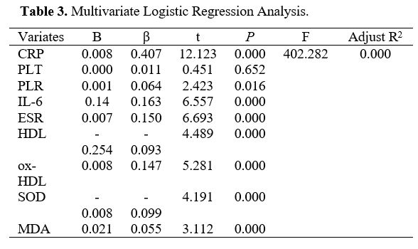
NCAL. The regression analysis was conducted to search for independent 
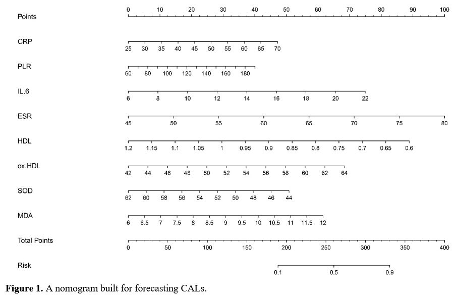
covariates for CALs related to OS and inflammation. The predictive 
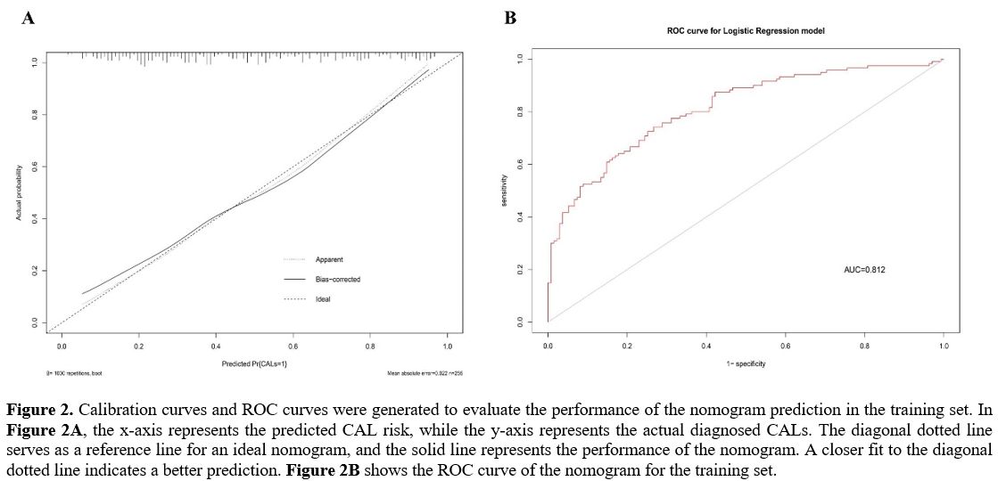
nomogram was structured according to these risk factors. The properties of the model were estimated using calibration and receiver operating characteristic curves.
Results
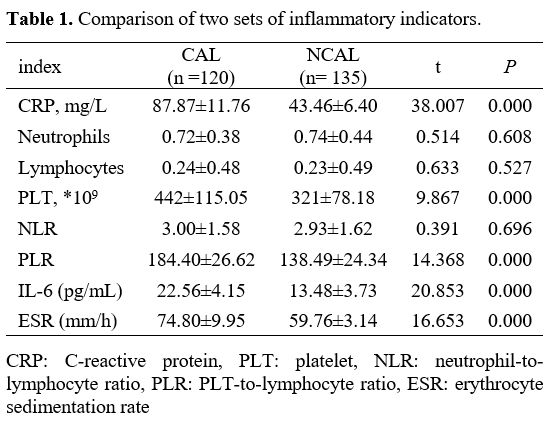
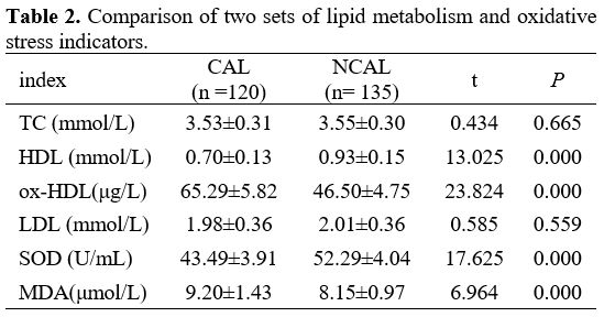
: The levels of CRP, IL-6, PLT count, ESR, ox-HDL, MDA, and PLR were more elevated in CAL patients with KD; interestingly, HDL and superoxide dismutase (SOD) were low in the CAL group. Ascension of CRP, IL-6, ESR, ox-HDL, MDA, and PLR, and diminution of HDL and SOD were considered independent risk factors. The nomogram constructed using these factors demonstrated a satisfactory calibration degree and discriminatory 
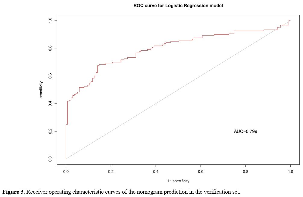
power, with an area under the curve of 0.812. In the verification set, the area under the curve was found to be 0.799.
Conclusion
: The model was established according to 8 OS and inflammation-associated risk factors bound up with CALs in KD sufferers. It may be a usable approach for early diagnosis of CALs in KD.


Introduction

Kawasaki Disease (KD) is the primary cause of posteriority heart disease in youngsters. It is characterized by acute angiitis of unexplained, which can result in coronary artery lesions (CAL) in 25% of untreated patients. In some cases, these lesions can lead to myocardial infarction and sudden death. However, this severe outcome only occurs in a sub-fraction of sufferers.[1] Presently, in the absence of a diagnostic kit available for KD, the clinical manifestation frequently poses challenges in distinguishing it from other febrile childhood diseases.[2,3] The consequences of an escape diagnosis of KD are significant, as prompt and adequate therapy is crucial in preventing the progression of CAL. The standard of care for acute KD is intravenous immune globulin (IVIG), which declines the proportion from 25% to 5%.[4,5] In addition, the lack of a diagnostic assay for KD poses challenges in identifying the condition and initiating IVIG treatment promptly, leading to increased morbidity and mortality associated with KD.
Inflammation and oxidative stress (OS) have a close relationship, with OS based on inflammation playing a significant part in the pathology of KD. The excessive production of reactive oxygen species (ROS) in the body leads to increased OS, initiating a continuous cycle of inflammatory reactions. This process is believed to contribute to the development of diffuse vasculitis during the acute phase.[6,7] To assess the degree of OS in vivo, researchers commonly measure ROS metabolites and blood biological antioxidant potential. There is evidence suggesting that these indices are highly expressed in patients with KD.[6] OS has been linked with various pathophysiological occurrences, such as the onset of vasculopathy, marked by an excess production of ROS.[8,9] The overabundance of ROS indicates endothelial impairment, resulting in the advancement of inflammation or malfunction in the coronary artery.[10] Hence, targeting oxidative stress has emerged as a potential strategy for managing cardiovascular conditions through the modulation of apoptosis and inflammation pathways.
Within the active regions of inflammation, various cells, such as vascular endothelial cells, are capable of generating ROS, active protein, and chemical medium, leading to OS. Additionally, OS irritates the NF-κB pathway and levels of cytokines and chemokines, further enhancing inflammation.[11-14] Therefore, inflammation and OS have close interaction and mutually amplify each other's effects. Some studies have established a significant relevance between the oxidative stress status in serum and the changes in levels of oxidized low-density lipoprotein, High-Density Lipoprotein (HDL), malondialdehyde, and F2-isoprostanes.[15,16] Studies have revealed that HDL may induce inflammation in macrophages. These inflammatory responses appear to be influenced by the passive removal of cholesterol from the cell membrane, triggering a cascade involving protein kinase C/NF-κB/STAT1-IRF1 signaling.[17] The researchers hypothesized a two-phase model of gene expression modulation, where an initial anti-inflammatory phase transitions to a pro-inflammatory phase.
Additionally, the inflammatory effects were found to be closely linked to cellular cholesterol levels, which govern the activation of IRE1a/ASK1/p38 MAPK and endoplasmic reticulum stress.[18] Adding oxidized high-density lipoprotein (ox-HDL) not only enhances NLRP3 expression but also triggers downstream cytokines and caspase-1, leading to the secretion of IL-1β and IL-18. Additionally, activation and induction effects appear to be dose-dependent. HDL presence reduces the expression of NLRP3 and inflammatory cytokines downstream.[19] Low-density lipoprotein (LDL) is a complex lipid-protein containing various types of lipids, such as triglycerides, phospholipids, and both free and esterified cholesterol. Scavenger receptors, such as CD36 and macrophage scavenger receptor 1 (MSR-1), play a crucial role in facilitating the absorption of Ox-LDL and work alongside toll-like receptors to convey inflammatory reactions in cells.[20] Ample proofs suggest that oxidative stress plays a significant part in regulating various aspects of thrombotic processes, such as platelet activation. It has been observed that oxidative stress can induce platelet hyperreactivity by reducing the availability of nitric oxide. Consequently, measuring oxidative stress levels could potentially aid in the early detection of asymptomatic individuals who are at risk of developing thrombosis.[21,22]
This study analyzed various common inflammatory indicators, along with serum lipids such as HDL, ox-HDL, LDL, and platelet (PLT) parameters. The objective was to explore the clinical significance of these indices in KD with CALs from the perspective of OS and identify additional treatment options for KD.


Materials and Methods

Sufferers. 510 Sufferers were randomly divided into two parts: training set (No.= 255) and verification set (No.=200). There is no significant difference in the proportion of patients with KD complicated with CALs between the two parts. The training set was categorized into two groups depending on whether it was with or without CAL, namely the CAL group (n=120) and the NCAL group (n=135). The study received approval from the Chengde Maternal and Child Health Care Hospital Ethics Committee, and the agreement was obtained from the parents or guardians of the children.
Diagnosis and Inclusion Criteria
. Both the diagnostic criteria for Kawasaki disease and Kawasaki disease CAL adhere to the diagnostic criteria published by the American Heart Association in 2017[23] and Evidence-based guidelines for the diagnosis and treatment of Kawasaki disease in Chinese children in 2023.[24] A fever that persists for 5 days or longer, without a definite end, and the existence of 2 or 3 of the 5 primary clinical signs: (1) absence of exudative conjunctival redness in both eyes; (2) Alterations in the lips and mouth, such as parchedness, redness, splitting, and redness of the lips, as well as wide redness of the mucous membranes in the lingonberry tongue and oropharynx; (3) Unexpected non-purulent enlargement of cervical lymph nodes (often diameter>1.5 cm); (4) Multifaceted rash, including single redness and puffiness near scars; (5) Transformations in the extremities, including the emergence of redness in the palms and toes, severe swelling in the hands and feet in the initial stage, and flaky peeling near the toenails in the healing phase. Simultaneously, definitive echocardiographic variations are observed when CAL is present, specifically (1) Z-value ≥ 2.0 for the left or right coronary artery or (2) coronary artery features that match the appropriate CAL criteria (internal diameter>3 mm in 4 mm in ≥ 5 years old, and/or adjacent internal diameter expansion ≥ 1.5 times or remarkably irregular lumen).
The study excluded patients with chronic cardiac insufficiency, cardiomyopathy, or organic heart disease, as well as those with other rheumatic immune diseases in combination. It also excluded children with recurrent KD and patients with incomplete laboratory data.
Erythrocyte Sedimentation Rate
. The automated erythrocyte sedimentation rate analyzer (CUBE 30 TOUCH, Monteriggioni (SI), Italia) utilizes a laser for illumination to analyze the minimal quantity of blood within the sedimentation tube. It dynamically assesses alterations in the aggregation and settling of red blood cells, consequently determining the results of the erythrocyte sedimentation rate.
Observation Indicators
. All patients should provide fasting blood samples before IVIG treatment and collect relevant blood index results.
ELISA
. Please refer to the following literature for specific operations.[25,26] Namely, the levels of superoxide dismutase (SOD), malondialdehyde (MDA), and interleukin-6 (IL-6) were detected using the ELISA kit (Spbio, Wuhan, China). The antigen was diluted to a concentration of 5 μg/mL and stayed overnight in the refrigerator. After that, the plate was cleaned three times with 1 × PBST washing solution (pH 7.2–7.4), with each wash lasting 3–5 minutes after the coating buffer was removed. A total of 250 μL of 1% BSA blocking buffer was put into each well. The wells were sealed and incubated at 35±2°C for 2h. Subsequently, the kit was washed three times using 1 × PBST for 3–5 minutes each time. The supernatants were diluted using a 1 × PBST at a ratio of 1:1,000, with 100 μL added to each well. The negative control was incubated at 37°C for 1.5 hours. A 1:1000 dilution of 1x PBST buffer was used to add 50 μL of the corresponding antibody to each well. The plate was then incubated at 37°C for 1 hour. Substrate solution was added to each well and incubated for 10 minutes at 37°C in the absence of light. After color development, 50 μL of termination solution was added to stop the reaction. The optical density at 450 nm was measured.
Statistical Analysis
. Data were administrated using SPSS 26 (SPSS, Inc., Chicago, IL, USA). The demographic characteristics, clinical manifestations, and laboratory data between the two groups were compared using the χ2 test, unpaired Student t-test, or Mann-Whitney U test. Important indicators identified from the univariate analysis were further analyzed using multivariate logistic regression analysis to determine independent risk factors of CALs. P<0.05 was considered statistically significant.

Results

Basic Information about Subjects. Information was collected from 255 patients, including 120 in CAL and 135 in NCAL. There are 85 male children in CAL, aged 0.8-4.25 years, with an average of (2.91 ± 1.06) years old, and 100 male children in NCAL, aged 0.7-5 years, with an average of (2.69 ± 1.01) years. There is no significant difference in gender and age.
Comparison of Two Sets of Inflammatory Indicators. As shown in Table 1, the levels of CRP, PLT, PLR, IL-6, and ESR in the CAL group were prominently more than that in the NCAL group. Interestingly, there is no difference in neutrophils, lymphocytes, and NLR between the two groups.


Table 1
Table 1. Comparison of two sets of inflammatory indicators.

Comparison of Two Sets of Lipid Metabolism and Oxidative Stress Indicators. As shown in Table 2, the levels of ox-HDL and MDA in the CAL group were dramatically more than that in the NCAL group; interestingly, the content of HDL and SOD in the CAL group was prominently low. There is no difference in TC and LDL between the two groups.

Table 2
Table 2. Comparison of two sets of lipid metabolism and oxidative stress indicators.

Multivariate Logistic Regression Analysis (MLRA). Statistically significant variables, including CRP, PLT, HDL, IL-6, ox-HDL, SOD, MDA, PLR, and ESR, were enrolled in the logistic regression analysis. Higher CRP, PLR, IL-6, MDA, ox-HDL, ESR, lower HDL, and SOD were independent factors for CALs with KD, whereas PLT failed to reach significant differences (Table 3).

Table 3
Table 3. Multivariate Logistic Regression Analysis.

Development and Evaluation of the Nomogram for Predicting CALs in KD. The nomogram was constructed using the eight independent risk factors identified through MLRA. (Figure 1). The calibration curve of the nomogram demonstrated good agreement in the CALs (Figure 2A). The area under the ROC curve (AUC) was 0.812 (95% CI, 0.76-0.865), and the sensitivity and specificity were 0.73 and 0.74, respectively (Figure 2B).
We further used the verification set to verify the model, which showed adequate discrimination (AUC, 0.799; 95% CI, 0.742-0.856). The sensitivity and specificity were 0.85 and 0.68, respectively (Figure 3).


Figure 1 Figure 1. A nomogram built for forecasting CALs.

Figure 2 Figure 2. Calibration curves and ROC curves were generated to evaluate the performance of the nomogram prediction in the training set. In Figure 2A, the x-axis represents the predicted CAL risk, while the y-axis represents the actual diagnosed CALs. The diagonal dotted line serves as a reference line for an ideal nomogram, and the solid line represents the performance of the nomogram. A closer fit to the diagonal dotted line indicates a better prediction. Figure 2B shows the ROC curve of the nomogram for the training set.

Figure 3 Figure 3. Receiver operating characteristic curves of the nomogram prediction in the verification set.


Discussion

Based on the results of the aforementioned OS-related indices, we have developed a novel nomogram model. This model incorporates 8 laboratory markers (CRP, IL-6, ESR, HDL, ox-HDL, SOD, and MDA) for predicting CALs in KD. The purpose of this model is to assist clinicians in detecting CALs at an earlier stage.
Since the initial discovery of Kawasaki disease, there has been a notable increase in its incidence rate. The combination of Kawasaki disease and CAL, along with its prognosis, continues to be a concern in the medical field. Numerous long-term follow-up studies conducted both domestically and internationally have revealed that Kawasaki disease, particularly when accompanied by CAL, serves as a hazard factor for ischemic heart disease.[27,28] The progression of CAL in KD is attributed to the impaired function of coronary endothelial cells resulting from the interplay between inflammation and oxidative stress.[29,30] The current lack of specific laboratory indicators for early diagnosis of Kawasaki disease and Kawasaki disease CAL poses a challenge. Cardiac ultrasound is only able to detect existing CAL and cannot identify previous endothelial cell dysfunction. Therefore, it is crucial to investigate the pathogenesis of Kawasaki disease further. This research is of great importance for the diagnosis, treatment, and prognosis of Kawasaki disease and Kawasaki disease CAL. The aim is to identify detection indicators that can predict early coronary artery damage.
CRP, an acute-phase blood protein, is yielded via the liver in response to inflammation and tissue damage. It is considered the most active marker of inflammation and plays a crucial role in inducing phagocytosis, cytokine production, and monocyte activation.[31] Previous studies have shown a significant increase in the mentioned factor during the acute phase of KD, especially in individuals with CALs, which yielded similar results to our investigation.[32,33] IL-6 is a widely used indicator of inflammation that directly reflects the state of a patient's immune system. Furthermore, IL-6 tends to change earlier than CRP following the onset of inflammatory reactions in the body. Consequently, the measurement of IL-6 levels can provide an accurate assessment of the patient's disease status.[34,35] In this study, it was observed that patients with CAL exhibited significantly elevated levels of IL-6, which aligns with findings from previous studies conducted by other researchers.[36,37]
NLR and PLR are inflammatory markers that are simple, convenient, and easily accessible.[38] Research has confirmed that children with KD combined with CAL have significantly elevated levels of NLR and PLR, which suggests that they could be potential risk factors for CAL. Additionally, this study confirms that PLR was elevated in the KD combined with the CAL group.[39] However, NLR is meaningless and may be the cause of the disease course. ESR is a widely used indicator for assessing inflammation levels. In children diagnosed with Kawasaki disease, the immune system becomes highly activated during the acute phase, leading to the production of significant amounts of fibrinogen and immunoglobulin, which contributes to an elevation in ESR. The rate of ESR is closely associated with the extent of inflammation and the potential injury to the coronary arteries within the body.[40] This study found that the results of ESN are consistent with other studies.[33,41]
HDL is widely recognized as a protective factor against cardiovascular diseases, including coronary atherosclerosis. It exhibits its antioxidant function through both direct and indirect effects. The direct antioxidant activity of HDL involves the elimination of lipid peroxides in the bloodstream, thereby preventing endothelial dysfunction, vascular damage, and the formation of CALs caused by continuous oxidative stress resulting from the oxidative modification of LDL. On the other hand, the indirect effect of HDL's antioxidant activity is manifested in its ability to facilitate cholesterol reverse transport and possess anti-inflammatory properties.[42,43] Ox-HDL is a product of lipid peroxidation formed after high-density lipoprotein is oxidized and modified, which has been proven to be one of the key factors in endothelial cell dysfunction. Endothelial injury is the initiating factor of vascular-related diseases. Ox-HDL has been confirmed to take major action on atherosclerosis, coronary heart disease, and chronic kidney disease.[44,45] SOD is a crucial component of the body's antioxidant enzyme and falls under the category of antioxidant metalloenzymes. It takes a major action on inhibiting the entire process of superoxide anion free radical disproportionation, leading to the production of oxygen and hydrogen peroxide. SOD helps maintain the balance between oxidation and oxidation in the body. MDA acts on the products of lipid peroxidation and can cause proteins to fuse with nucleic acids, resulting in cytotoxicity. High concentrations of MDA attacks can lead to pathological changes in cells, increasing the risk of oxidative stress. Therefore, clinical detection of SOD and MDA levels can reflect the risk of oxidative stress reactions in KDs, and that can further aid in developing preventive measures against potential risks. In our study, we observed that HDL and SOD levels were memorably abated, and Ox-HDL and MDA levels were memorably augmented in KD with CALs; moreover, the decrease in HDL and SOD or increase in MDA and Ox-HDL was a factor for CALs, which is consistent with previous studies.[39,46]
In this study, the CAL group exhibited a significant increase in PLT compared to the NCAL group. Previous studies have yielded inconsistent findings regarding the association between PLT and risk for CALs. While some studies have suggested a higher risk of CALs with lower PLT counts, others have reported a higher risk with higher PLT counts. We found that patients with abnormally high PLT at admission were more likely to develop CALs. Our results partially support the findings of their study.[47,48]

Conclusions

This study found significant differences in various indicators that indirectly reflect the level of OS and inflammation between CAL and NCAL groups. This paper also confirmed that a nomogram can effectively predict the risk of CALs using CRP, IL-6, ESR, HDL, ox-HDL, SOD, and MDA. These findings suggest that when hospital laboratory resources are limited, standard detection indices such as HDL, ox-HDL, and ESR can be used to forecast the level of OS with KD and identify the risk of CALs early. Additionally, the study highlights the role of OS in CALs secondary to KD and suggests that early antioxidant therapy could be an important adjuvant treatment to improve prognosis.

Ethics Approval and Consent to Participate

This study was approved by the Medical Ethics Committee of Chengde Maternal and Child Health Care Hospital. All methods were conducted in accordance with relevant guidelines and regulations. All procedures were performed in accordance with the ethical standards laid down in the 1964 Declaration of Helsinki and its later amendments. Informed consent was obtained from their legal guardian (s).

Authors Contributions

Y. L. analyzed data and writing. S. W. L. participated in the collection of data and experimental operations. H. B. Z. participated in design testing and supervision testing. All authors read and approve the manuscript version final

References   

  1. BAJOLLE F. [Diagnosis and management of Kawasaki disease] [J]. La Revue du praticien, 2023, 73(2): 179-86.  
  2. ABRAHAM D, KALYANASUNDARAM S, KRISHNAMURTHY K. Refractory Kawasaki Disease-a Challenge for the Pediatrician [J]. SN comprehensive clinical medicine, 2021, 3(3): 855-60. https://doi.org/10.1007/s42399-021-00775-w PMid:33532696 PMCid:PMC7843002  
  3. LAM J Y, SHIMIZU C, TREMOULET A H, et al. A machine-learning algorithm for diagnosis of multisystem inflammatory syndrome in children and Kawasaki disease in the USA: a retrospective model development and validation study [J]. The Lancet Digital Health, 2022, 4(10): e717-e26.
  4. BRODERICK C, KOBAYASHI S, SUTO M, et al. Intravenous immunoglobulin for the treatment of Kawasaki disease [J]. Cochrane Database of Systematic Reviews, 2023, 1(1): Cd014884. https://doi.org/10.1002/14651858.CD014884.pub2 PMid:36695415 PMCid:PMC9875364  
  5. LI Z, CAI J, LU J, et al. The therapeutic window of intravenous immunoglobulin (IVIG) and its correlation with clinical outcomes in Kawasaki disease: a systematic review and meta-analysis [J]. Italian Journal of Pediatrics, 2023, 49(1): 45. https://doi.org/10.1186/s13052-023-01451-6 PMid:37038188 PMCid:PMC10088291  
  6. HARA T, YAMAMURA K, SAKAI Y. The up-to-date pathophysiology of Kawasaki disease [J]. Clinical & Translational Immunology, 2021, 10(5): e1284. https://doi.org/10.1002/cti2.1284 PMid:33981434 PMCid:PMC8109476  
  7. YAHATA T, HAMAOKA K. Oxidative stress and Kawasaki disease: how is oxidative stress involved from the acute stage to the chronic stage? [J]. Rheumatology (Oxford), 2017, 56(1): 6-13. https://doi.org/10.1093/rheumatology/kew044 PMid:27094597  
  8. AMPEM P T, SMEDLUND K, VAZQUEZ G. Pharmacological evidence for a role of the transient receptor potential canonical 3 (TRPC3) channel in endoplasmic reticulum stress-induced apoptosis of human coronary artery endothelial cells [J]. Vascular Pharmacology, 2016, 76: 42-52. https://doi.org/10.1016/j.vph.2015.07.011 PMid:26215710 PMCid:PMC4706806  
  9. XING Y J, LIU B H, WAN S J, et al. A SGLT2 Inhibitor Dapagliflozin Alleviates Diabetic Cardiomyopathy by Suppressing High Glucose-Induced Oxidative Stress in vivo and in vitro [J]. Frontiers in pharmacology, 2021, 12: 708177. https://doi.org/10.3389/fphar.2021.708177 PMid:34322029 PMCid:PMC8311522  
  10. XU M, QI Q, MEN L, et al. Berberine protects Kawasaki disease-induced human coronary artery endothelial cells dysfunction by inhibiting of oxidative and endoplasmic reticulum stress [J]. Vascular Pharmacology, 2020, 127: 106660. https://doi.org/10.1016/j.vph.2020.106660 PMid:32070767  
  11. ZHANG Z, ZHAO L, ZHOU X, et al. Role of inflammation, immunity, and oxidative stress in hypertension: New insights and potential therapeutic targets [J]. Frontiers in Immunology, 2022, 13: 1098725. https://doi.org/10.3389/fimmu.2022.1098725 PMid:36703963 PMCid:PMC9871625  
  12. CAO Y Y, WANG Z, WANG Z H, et al. Inhibition of miR-155 alleviates sepsis-induced inflammation and intestinal barrier dysfunction by inactivating NF-κB signaling [J]. International immunopharmacology, 2021, 90: 107218. https://doi.org/10.1016/j.intimp.2020.107218 PMid:33296782  
  13. SUN S, ZHANG J, LI H, et al. Anti-inflammatory activity of the water extract of Chloranthus serratus roots in LPS-stimulated RAW264.7 cells mediated by the Nrf2/HO-1, MAPK and NF-κB signaling pathways [J]. Journal of ethnopharmacology, 2021, 271: 113880. https://doi.org/10.1016/j.jep.2021.113880 PMid:33508367  
  14. SUN X, ZHENG Y, TIAN Y, et al. Astragalus polysaccharide alleviates alcoholic-induced hepatic fibrosis by inhibiting polymerase I and transcript release factor and the TLR4/JNK/NF-κB/MyD88 pathway [J]. Journal of ethnopharmacology, 2023, 314: 116662. https://doi.org/10.1016/j.jep.2023.116662 PMid:37207880  
  15. KARABACAK M, UYSAL B A, TURKDOGAN A K. Alteration in serum oxidative stress balance in patients with different circulating high-density lipoprotein cholesterol levels [J]. The Portuguese Journal of Cardiology, 2022, 41(10): 833-9. https://doi.org/10.1016/j.repc.2021.06.022 PMid:36210589  
  16. SIMANTIRIS S, PAPASTAMOS C, A S A, et al. Oxidative stress biomarkers in coronary artery disease [J]. Current Topics in Medicinal Chemistry, 2023. https://doi.org/10.2174/1568026623666230502140614 PMid:37138428  
  17. SHI L, JIANG C, XU H, et al. Hyperoside ameliorates cerebral ischaemic-reperfusion injury by opening the TRPV4 channel in vivo through the IP(3)-PKC signalling pathway [J]. Pharmaceutical biology, 2023, 61(1): 1000-12. https://doi.org/10.1080/13880209.2023.2228379 PMid:37410551 PMCid:PMC10327524  
  18. NAZIR S, JANKOWSKI V, BENDER G, et al. Interaction between high-density lipoproteins and inflammation: Function matters more than concentration! [J]. Advanced Drug Delivery Reviews, 2020, 159: 94-119. https://doi.org/10.1016/j.addr.2020.10.006 PMid:33080259  
  19. LI W L, HUA L G, QU P, et al. NLRP3 inflammasome: a novel link between lipoproteins and Atherosclerosis [J]. Archives of Medical Science (AMS), 2016, 12(5): 950-8. https://doi.org/10.5114/aoms.2016.61356 PMid:27695484 PMCid:PMC5016581  
  20. OBERMAYER G, AFONYUSHKIN T, BINDER C J. Oxidized low-density lipoprotein in inflammation-driven thrombosis [J]. The Journal of Thrombosis and Haemostasis (JTH), 2018, 16(3): 418-28. https://doi.org/10.1111/jth.13925 PMid:29316215  
  21. FUENTES E, MOORE-CARRASCO R, DE ANDRADE PAES A M, et al. Role of Platelet Activation and Oxidative Stress in the Evolution of Myocardial Infarction [J]. Journal of Cardiovascular Pharmacology and Therapeutics (JCPT), 2019, 24(6): 509-20. https://doi.org/10.1177/1074248419861437 PMid:31280622  
  22. KHODADI E. Platelet Function in Cardiovascular Disease: Activation of Molecules and Activation by Molecules [J]. Cardiovascular Toxicology, 2020, 20(1): 1-10. https://doi.org/10.1007/s12012-019-09555-4 PMid:31784932  
  23. Correction to: Diagnosis, Treatment, and Long-Term Management of Kawasaki Disease: A Scientific Statement for Health Professionals From the American Heart Association [J]. Circulation, 2019, 140(5): e181-e4. https://doi.org/10.1161/CIR.0000000000000703  
  24. [Evidence-based guidelines for the diagnosis and treatment of Kawasaki disease in children in China (2023)] [J]. Zhongguo dang dai er ke za zhi = Chinese journal of contemporary pediatrics, 2023, 25(12): 1198-210.  
  25. CAO Y-Y, WANG Z, WANG Z-H, et al. Inhibition of miR-155 alleviates sepsis-induced inflammation and intestinal barrier dysfunction by inactivating NF-κB signaling [J]. International immunopharmacology, 2021, 90: 107218. https://doi.org/10.1016/j.intimp.2020.107218 PMid:33296782  
  26. ZHANG Y X, ZHANG X T, LI H J, et al. Antidepressant-like effects of helicid on a chronic unpredictable mild stress-induced depression rat model: Inhibiting the IKK/IκBα/NF-κB pathway through NCALD to reduce inflammation [J]. International immunopharmacology, 2021, 93: 107165. https://doi.org/10.1016/j.intimp.2020.107165 PMid:33578182  
  27. SEKI M, MINAMI T. Kawasaki Disease: Pathology, Risks, and Management [J]. Vascular Health and Risk Management, 2022, 18: 407-16. https://doi.org/10.2147/VHRM.S291762 PMid:35711626 PMCid:PMC9196282  
  28. THOMAS K N, AGGARWAL A. Childhood rheumatic diseases: bites not only the joint, but also the heart [J]. Clinical Rheumatology, 2023: 1-13. https://doi.org/10.1007/s10067-023-06621-9 PMid:37160484 PMCid:PMC10169151  
  29. WANG Y, HU J, LIU J, et al. The role of Ca(2+)/NFAT in Dysfunction and Inflammation of Human Coronary Endothelial Cells induced by Sera from patients with Kawasaki disease [J]. Scientific Reports, 2020, 10(1): 4706. https://doi.org/10.1038/s41598-020-61667-y PMid:32170198 PMCid:PMC7069934  
  30. XU M, QI Q, MEN L, et al. Berberine protects Kawasaki disease-induced human coronary artery endothelial cells dysfunction by inhibiting of oxidative and endoplasmic reticulum stress [J]. Vascular Pharmacology, 2020, 127: 106660. https://doi.org/10.1016/j.vph.2020.106660 PMid:32070767  
  31. AGRAWAL A. CRP after 2004 [J]. Molecular Immunology, 2005, 42(8): 927-30. https://doi.org/10.1016/j.molimm.2004.09.028 PMid:15829284 PMCid:PMC3833354  
  32. SHUAI S, ZHANG H, ZHANG R, et al. Prediction of coronary artery lesions based on C-reactive protein levels in children with Kawasaki Disease: a retrospective cohort study [J]. The Jornal de Pediatria, 2023. https://doi.org/10.1016/j.jped.2023.02.005 PMid:36977496 PMCid:PMC10373155  
  33. YANG Y, HU X. The Predictive Values of MMP-9, PLTs, ESR, and CRP Levels in Kawasaki Disease with Cardiovascular Injury [J]. Evidence-Based Complementary and Alternative Medicine (eCAM), 2022, 2022: 6913315. https://doi.org/10.1155/2022/6913315 PMid:36159562 PMCid:PMC9492360  
  34. DEL GIUDICE M, GANGESTAD S W. Rethinking IL-6 and CRP: Why they are more than inflammatory biomarkers, and why it matters [J]. Brain, Behavior, and Immunity, 2018, 70: 61-75. https://doi.org/10.1016/j.bbi.2018.02.013 PMid:29499302  
  35. HEINRICH P C, BEHRMANN I, HAAN S, et al. Principles of interleukin (IL)-6-type cytokine signalling and its regulation [J]. Biochemical Journal, 2003, 374(Pt 1): 1-20. https://doi.org/10.1042/bj20030407 PMid:12773095 PMCid:PMC1223585  
  36. CHANG L, YANG H W, LIN T Y, et al. Perspective of Immunopathogenesis and Immunotherapies for Kawasaki Disease [J]. Frontiers in Pediatrics, 2021, 9: 697632. https://doi.org/10.3389/fped.2021.697632 PMid:34350146 PMCid:PMC8326331  
  37. LI Y, ZHENG Q, ZOU L, et al. Kawasaki disease shock syndrome: clinical characteristics and possible use of IL-6, IL-10 and IFN-γ as biomarkers for early recognition [J]. Pediatric Rheumatology, 2019, 17(1): 1. https://doi.org/10.1186/s12969-018-0303-4 PMid:30611297 PMCid:PMC6321686  
  38. KOSIDŁO J W, WOLSZCZAK-BIEDRZYCKA B, MATOWICKA-KARNA J, et al. Clinical Significance and Diagnostic Utility of NLR, LMR, PLR and SII in the Course of COVID-19: A Literature Review [J]. Journal of Inflammation Research, 2023, 16: 539-62. https://doi.org/10.2147/JIR.S395331 PMid:36818192 PMCid:PMC993057  
  39. WU R, JIANG W, SUN Y, et al. Indicators of Oxidative Stress in the Prediction of Coronary Artery Lesions in Patients With Kawasaki Disease [J]. Journal of clinical rheumatology: practical reports on rheumatic & musculoskeletal diseases, 2023, 29(3): 126-31. https://doi.org/10.1097/RHU.0000000000001925 PMid:36730421
  40. LAPIĆ I, PADOAN A, BOZZATO D, et al. Erythrocyte Sedimentation Rate and C-Reactive Protein in Acute Inflammation [J]. The American Journal of Clinical Pathology (AJCP), 2020, 153(1): 14-29. https://doi.org/10.1093/ajcp/aqz142 PMid:31598629  
  41. ZHOU C, ZHAO Y, WANG X, et al. Laboratory parameters between multisystem inflammatory syndrome in children and Kawasaki disease [J]. Pediatric Pulmonology (PPUL), 2021, 56(12): 3688-98. https://doi.org/10.1002/ppul.25687 PMid:34583417  
  42. GANJALI S, GOTTO A M, JR., RUSCICA M, et al. Monocyte-to-HDL-cholesterol ratio as a prognostic marker in cardiovascular diseases [J]. The Journal of Cellular Physiology, 2018, 233(12): 9237-46. https://doi.org/10.1002/jcp.27028 PMid:30076716  
  43. SHOAR S, IKRAM W, SHAH A A, et al. Non-high-density lipoprotein (non-HDL) cholesterol in adolescence as a predictor of atherosclerotic cardiovascular diseases in adulthood [J]. Reviews in Cardiovascular Medicine (RCM), 2021, 22(2): 295-9. https://doi.org/10.31083/j.rcm2202037 PMid:34258898  
  44. MATSUNAGA T, IGUCHI K, NAKAJIMA T, et al. Glycated high-density lipoprotein induces apoptosis of endothelial cells via a mitochondrial dysfunction [J]. Biochemical and Biophysical Research Communications, 2001, 287(3): 714-20. https://doi.org/10.1006/bbrc.2001.5625 PMid:11563854  
  45. SUN J T, CHEN Y Y, MAO J Y, et al. Oxidized HDL, as a Novel Biomarker for Calcific Aortic Valve Disease, Promotes the Calcification of Aortic Valve Interstitial Cells [J]. Journal of Cardiovascular Translational Research (JCTR), 2019, 12(6): 560-8. https://doi.org/10.1007/s12265-019-09903-3 PMid:31367900  
  46. LEBRANCHU Y, MALVY D, RICHARD M J, et al. Kawasaki disease and oxidative metabolism [J]. Clinica Chimica Acta, 1990, 187(3): 193-8. https://doi.org/10.1016/0009-8981(90)90104-Z PMid:2108832
  47. AE R, ABRAMS J Y, MADDOX R A, et al. Platelet Count Variation and Risk for Coronary Artery Abnormalities in Kawasaki Disease [J]. The Pediatric Infectious Disease Journal, 2020, 39(3): 197-203. https://doi.org/10.1097/INF.0000000000002563 PMid:31851145
  48. HUIJUAN K, YAPING D, BO W, et al. Combined IFN-β and PLT Detection Can Identify Kawasaki Disease Efficiently [J]. Frontiers in Pediatrics, 2021, 9: 624818. https://doi.org/10.3389/fped.2021.624818 PMid:33968843 PMCid:PMC8100027