Recent Advances in the Definition of the Molecular Alterations Occurring in Multiple Myeloma 

Ugo Testa1, Elvira Pelosi1, Germana Castelli1 and Giuseppe Leone2.

1 Hematology, Department of Translational and Precision Medicine; "Sapienza" University of Rome, Italy.
2 Resident Doctor at the Department of Hematology, University “Sapienza” of Rome, Rome, Italy.

Correspondence to:  Ugo Testa. E-mail: ugo.testa@iss.it

Published: July 01, 2024
Received: June 13, 2024
Accepted: June 19, 2024
Mediterr J Hematol Infect Dis 2024, 16(1): e2024062 DOI 10.4084/MJHID.2024.062

This is an Open Access article distributed under the terms of the Creative Commons Attribution License
(
https://creativecommons.org/licenses/by-nc/4.0), which permits unrestricted use, distribution, and reproduction in any medium, provided the original work is properly cited.

Abstract

Multiple myeloma (MM) is a disorder of the monoclonal plasma cells and is the second most common hematologic malignancy. MM initiation and 
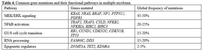
progression are dependent upon complex genomic abnormalities. The 
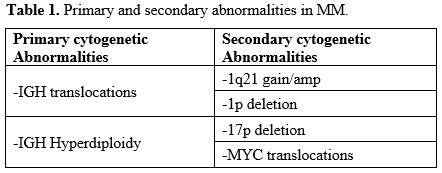
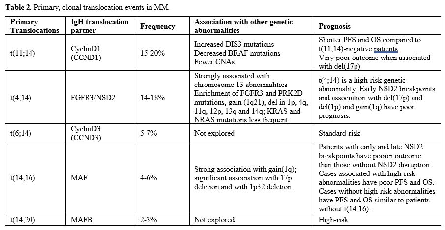
current pathogenic model of MM includes two types of primary events, 
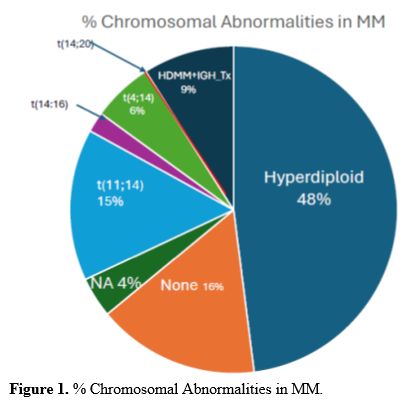
represented by chromosome translocations or chromosome number alterations resulting in hyperdiploidy. These primary molecular events are observed both in MM and in monoclonal gammopathy, its premalignant 
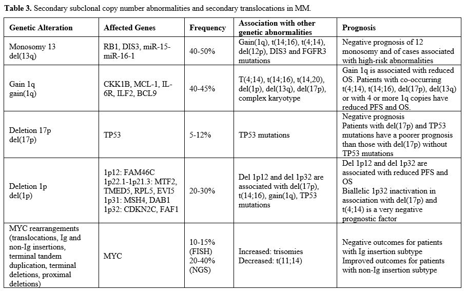
precursor. Subsequent genetic events allow the progression of monoclonal gammopathy to MM and, together with primary events, contribute to the genetic complexity and heterogeneity of MM.
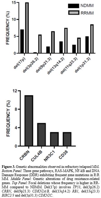
Newer therapies have considerably improved patient outcomes; however, MM remains an incurable disease and most patients experience multiple relapses.
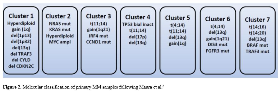
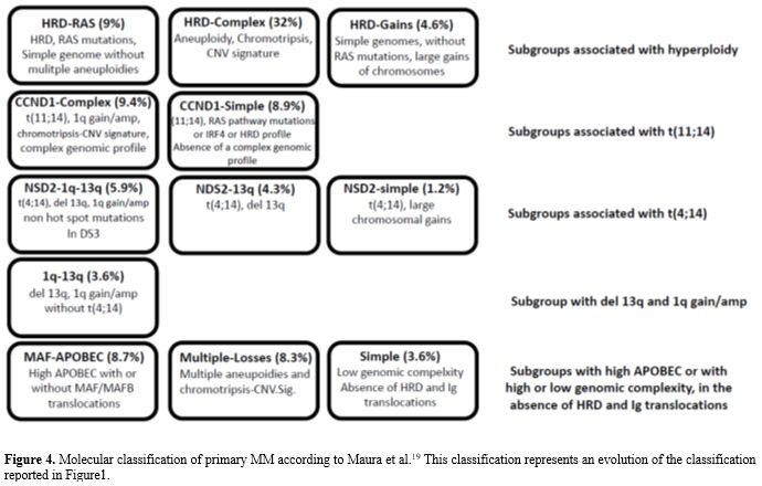
The dramatic progresses achieved in the analysis of the heterogeneous molecular features of different MM patients allowed a comprehensive molecular classification of MM and the definition of an individualized prognostic model to predict an individual MM patient’s 
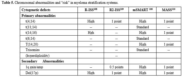
response to different therapeutic options. Despite these progresses, prognostic models fail to identify a significant proportion of patients destined to early relapse. Treatment strategies are increasingly. Based on disease biology, trials are enriched for high-risk MMs, whose careful definition and categorization requires DNA sequencing studies.


Introduction

Multiple myeloma (MM) is a malignant disease of mature B-cell lineage, characterized by the proliferation and accumulation of plasma cells (PCs) in bone marrow with consequent production of a monoclonal antibody. The development of MM is a multistep process through three different tumor stages: (i) an asymptomatic premalignant condition, known as monoclonal gammopathy of undermined significance (MGUS), characterized by the presence in the bone marrow of few abnormal plasma cells and of a monoclonal (M) protein instead of normal antibodies; (ii) a more advanced condition, called smouldering multiple myeloma (SMM), characterized by a higher serum level of M protein and a higher percentage of abnormal PCs in BM; about 50% of patients with SMM show a progressive increase of monoclonal protein and develop MM.[1,2]

Genetic Alteration in Multiple Myeloma

Cytogenetic studies have shown that MM can be split into cases with primary immunoglobulin translocations and cases with hyperdiploidia with trisomies of the odd-manner chromosomes: the most frequent translocations are t(11;14), t(4;14), t(14;16), t(14;20) and t(6;14); the most frequent copy number gains and losses are del13q, 1q1, del14q, del6q, del1p and del17p.[3] (Table 1 and Table 2)

Table 1 Table 1. Primary and secondary abnormalities in MM.

Table 2 Table 2. Primary, clonal translocation events in MM.


Primary Genetic Abnormalities in MM

Hyperdiploidy. Hyperdiploid MMs are characterized by recurrent chromosome gains, called chromosomal numerical abnormality (CNA). These tumors have 48-75 chromosomes, usually with extra copies of three more specific chromosomes. Hyperdiploid tumors rarely (about 10%) have a primary IgH translocation. The molecular mechanism responsible for development of hyperdiploidy is seemingly related to errors in chromosome segregation during the highly proliferative germinal centre phase of plasma cell ontogeny, as a result of a single catastrophic mitotic event or of multiple aberrant segregation events.[4
In hyperdiploid MMs gains in chromosomes 19 (95%), 15 (90%) and 9 (90%) are the most frequent events, followed by gains in other chromosomes, such a 5, 11, 3, 7 and 21; del13 is the most common deletion event, observed in 37% of these tumors; the majority of events occurring at lower frequencies in these tumors are deletions; the large majority (>90%) of hyperdiploid tumors had concurrent gains in at least two of the three chromosomes most frequently associated with trisomies.[5] In the non-hyperdiploid MMs, gain of chromosome 11 (92% of patients) and del 13 (99%) and gain of 1q are the most frequent clonal events, suggesting an important role in the early stages of disease.[5]
A chronological reconstruction of aneuploidies acquisition in hyperdiploid patients showed that: in individual patients pronounced changes in their karyotype were observed over time, including chromosome gains; in 13/18 hyperdiploid patients cumulative acquisition of copy-number gains was observed, while in the remaining  5 patients trisomies were acquired in one single time window.[6]   
The group of hyperdiploid MMs is heterogeneous for the variable association with additional genetic alterations. Hyperdiploid MMs can be subdivided into two subgroups according to the presence or not of concomitant gain(11q25); tumors lacking gain(11q25) characterized by gain(1q).[7]
Barilà and coworkers have defined two subsets of hyperdiploid MM patients, one characterized by 5 trisomies and defined as T-HRD and the other one by <5 trisomies and defined as N-HRD; T-HRD were characterized by a better outcome than N-HRD patients (mOS 57 vs 32 months).[8] T-HRD MMs were associated with low rates of FISH alterations compared to N-HRD.[8]
MM patients with hyperdiploidy have a better survival than those without hyperdiploidy when treated with novel anti-myeloma agents [9]. However, the presence of hyperdiploidy cannot ameliorate the negative prognostic impact of concurrent high-risk cytogenetic abnormalities.[9] Samur and coworkers, through whole genome sequencing identified a subgroup of MM patients (17% of total), 90% hyperdiploid, with low DNA damage (low genomic scar score with chromosome 9 gain), with frequent NRAS mutations, associated with very good outcome (100% overall survival at 69 months).[10]
Although it is currently assumed that the two founding events in MM pathogenesis, hyperdiploidy and IgH translocations are mutually exclusive, it was observed that in 4% of newly diagnosed MM patients hyperdiploidy and IgH translocations occur concurrently.[11]
IgH Translocations. IgH translocations have an important oncogenic effect, placing oncogenes under the control of strong enhancers (Ig heavy chain (IgH) loci). The five most recurrent IgH translocations observed in MM are represented by the: (i) translocation to the long arm of chromosome 11 t(11;14) involving cyclin D1 (CCDN1), observed in about 16% of cases; translocation to the short arm of chromosome 4 t(4;14) involving FGFR3/NSD2, observed in about 15% of cases; translocation to the short arm of chromosome 6 t(6;14) involving Cyclin D3 (CCDN3), occurring in about 6% of cases; translocation to the long arm of chromosome 16 t(16;14) involving MAF, occurring in about 5% of cases; long arm of chromosome 20 t(14;20) involving MAFB, occurring in about 2% of cases [12]. (Table 2) When present, these translocations are always clonal events.
T(11;14). Translocations dysregulating cyclin D expression are the most recurrent type of IgH translocations and involve cyclin D1 t(11;14), cyclin D3 t(6;14) and cyclin D2 t(12;14) and lead to increased expression of the corresponding cyclin genes. However, a dysregulated and/or increased expression of cyclin D1, D2 or D3 is a common feature not only of these IgH translocations, but also of other IgH translocations, as well as of hyperdiploid MMs.[12] While cyclin D1 and D3 overexpression is directly related to translocations that dysregulate CCND1 (11q13) or CCND3 (6p21), cyclin D2 overexpression is either directly induced by translocations that affect CCND2 (12p13)[13,14] or by translocations affecting MAF (16q23) or MAFB (20q11) transcription factors that target CCND2.[12,15]
Some evidences suggest that t(11;14) could represent an unique MM subset with peculiar biological properties, as evidenced by higher levels of the antiapoptotic protein BCL-2 and frequent expression of the B-cell lineage protein CD20 [16]. Characterization of a large cohort of t(11;14)-positive MM patients by NGS showed that these patients have a differentiated genetic architecture, compared to t(11;14)-negative patients, characterized by fewer CNAs associated with increased genomic stability, but increased rates of DIS3 mutations and decreased rates of BRAF mutations.[17] Avet-Loiseau confirmed the presence of a markedly increased frequency of DIS3 mutations and a decreased frequency of BRAF mutations in t(11;14) MM.[18]
The prognosis of t(11;14) can be explained by its binary genomics, i.e., cases with very few other lesions and cases with high-risk genetic abnormalities behave differently.[19]
Ziccheddu et al. have analyzed 514 newly diagnosed MM and showed that t(11;14) and chr(1q)gain/amps predicted differential expression of the BCL-2 axis and response to Venetoclax.[20] The BCL2/BCL2L1 ratio was high in t(11;14) setting, explaining the positive effect of Venetoclax in this subgroup; In contrast, chr(1q)gain/amps display a low BCL2/BCL2L1 ratio and lead to Venetoclax resistance through MCL1 overexpression.[20]
The oral BCL-2 inhibitor Venetoclax has shown promising efficacy in patients with t(11;14) MM patients, both a single-agent and in combination. Several ongoing trials are exploring Venetoclax in t(11;14) MM patients.[21-25] However, in relapsed/refractory MM patients the phase III placebo-controlled BELLINI trial failed to show superior outcomes from Venetoclax in combination with bortezomib and dexamethasone compared to placebo plus bortezomib and dexamethasone.[21] MCL1 and BCL2L1 copy number gains and structural rearrangements were linked to Venetoclax resistance in t(11;14) MM.[26]
T(4;14). T(4;14) is the second most-common translocation, occurring in about 15% of newly diagnosed MMs. This is an example of an IgH translocation resulting in the dysregulation of two different genes with oncogenic potential: FGFR3 and MMSET (named NSD2). The t(4;14) was strongly associated with chromosome 13 abnormalities.[27]
Keats et al. have used a RT-PCR-specific assay to detect hybrid IgH-NSD2 transcript and observed a frequency of about 15% in MM and about 2% in MGUS; the presence of t(4;14) was predictive of poor response to first line chemotherapy and reduced OS.[28] The analysis of 67 t(4;14) patients showed in 10% of cases FGFR3 mutations, in 44% FGFR3 overexpression without FGFR3 mutations and in 28% absent FGFR3 expression; adverse prognosis was restricted only to patients with FGFR3 mutations.[29]
Analysis of the genomic landscape of t(4;14) newly diagnosed MM showed enrichment of mutations in FGFR3 (38%) and PRK2D (7%), amplifications of 1q21 and deletions in 1p, 4q, 11q, 12p, 13q and 14q; KRAS and NRAS mutations are less frequent in t(4;14) than in non-t(4;14) MMs.[30]
Walker et al. have performed a whole sequencing study and have analyzed the IgH locus breakpoints and identified breakpoints either of the NSD2 gene or within the coding sequence of this gene.[31] Only patients with a breakpoint within the NSD2 gene and downstream the translation start site (identified as late disruptions, corresponding to 31% of these patients) have a worse overall survival; in contrast, patients with a breakpoint between the transcription and the translation start site (identified as early disruption, corresponding to 23.5% of these patients) and upstream (identified as no disruption, corresponding 45.5% of these patients) of the NSD2 gene exhibited progressively longer survival.[32]
Geng and coworkers have analyzed the impact of t(4;14) translocation in a group of 606 ND MM patients, including 108 t(4;14) cases.[33] Median OS (56.2 vs 87.3 months) and PFS (25.7 vs 37.6 months) were significantly shorter in patients with t(4;14) than in those without this cytogenetic abnormality.[33] Among the patients with t(4;14), 26.9% had t(4;14) alone, 59.3% had t(4;14) with gain (1q21), 13.9% had t(4;14) with both gain (1q21) and del(17p): patients with t(4;14) alone have an OS comparable to the rest of MM patients, while those with t(4;14) in association with amp (1q21) and del (17p) have a reduced OS.[33] Thus, t(4;14) alone, in the absence of gain (1q21) and del (17p) have a reduced OS, as confirmed by another study.[34] In MM, amplification or gain of chromosome 1q (1q
+) can involve the whole long arm of chromosome 1 or only specific cytobands such as 1q21, 1q22 or 1q23.3. However, it is important to note that, while the survival of double-hit t(4;14) and 1q21+ or 1q23+ or both is lower than that of t(4;14) alone, the survival of double-hit MM was similar to that displayed by 1q21+ or 1q23+ alone.[34]
T(14;16) and t(14;20). The translocations (14;16) and (14;20) are less common (3-5% and 1-2%, respectively)  involve the IGH locus and the oncogene c-MAF and MAFB, respectively. MAF induces expression of CCND2 and integrin B7, two events that stimulate MM cell proliferation. Next generation sequencing studies on 5141 newly diagnosed MM have identified 169 (3.3%) t(14;16) cases whose characterization showed a high association with high-risk abnormalities: gain/amplification of 1q was observed in 69% of patients with t(14;16) compared to 29% in those without t(14;16); deletion 1p32 was detected in 20.7% of patients with t(14;16) compared to 8.5% in those without t(14;16); biallelic 1p32 deletion was observed in 4.7% of patients with t(14;16) compared to 1.8% in those without t(14;16); 17p deletion was observed in 22.5% of patients with t(14;16) compared to 8.7% in those without t(14;16); biallelic TP53 inactivation was observed in 8.9% of patients with t(14;16) compared to 3.1% in those without t(14;16); TP53 mutations were detected in 14.2% of cases with t(14;16) compared to 5.5% in those without t(14;16).[32] The t(14;16) has not any prognostic impact if isolated (but numbers are very small). In contrast, its interaction with another prognostic lesion can lead to an aggressive disease.[35] Clinical data showed that patients with t(14;16) have shorter mPFS (14.3 months) and mOS (61.3 months) compared to those without t(14;16) who have mPFS of 43.9  months and mOS of 128.8 months; However, the shorter mPFS and mOS observed is due to the association with high-risk abnormalities.[35] Cyclin D2 protein was observed in all the cases bearing t(14;16), but in only 24% of those bearing t(4;14) 1q gains.[36]
In MM patients with t(14;16) and t(14;20) are frequent APOBEC ("apolipoprotein B mRNA editing enzyme, catalytic polypeptide") family mutational signatures (SBS2 and SBS13); patients with this signature have an increased mutational load and poor outcomes.[34] Overexpression of MAF and MAFB expression results in increased APOBEC3B and APOBEC4 expression, and consequent induction of DNA mutations.[37] A recent whole exome sequencing study carried out in 726 MM patients identified APOBEC mutational activity in 57.5% of these patients; however, only 6.6% MM patients were defined as hyper APOBEC, the majority of them (74%) being t(14;16)-positive.[38]

Secondary Cytogenetic Abnormalities

Subclonal copy number alterations. The most frequent subclonal CNAs observed in MM include gain of the long arm of chromosome 1 (gain 1q), deletion of the long arm of chromosome 13 (del(13q), deletion of the long arm of chromosome 14 (del(14q)), deletion of the short arm of chromosome 17 (del(17p)) and deletion of the short arm of chromosome 1 (del(1p). (Table 3)

Table 3
Table 3. Secondary subclonal copy number abnormalities and secondary translocations in MM.

Gain(1q). Gain 1q occurs in about 40% of patients and is preferentially associated with other cytogenetic abnormalities compared to MMs without gain 1q, involving an higher frequency of t(4;14) and t(14;16), of del(1p), del(17p) and particularly del(13q); furthermore, MMs with gain(1q) have an higher frequency of complex karyotype compared to those without gain(1q).[39]
The majority of studies have shown a negative impact of gain(1q) on PFS and OS.[7,36,40]
Several studies have evaluated the outcomes of MM patients with 1q gain who received auto-HSCT. In this context, a study from Mayo Clinic, including 155 MM patients undergoing upfront auto-HSCT, showed a shorter OS in patients with 1q+ compared to patients without this genetic abnormality.[41] In a subgroup of the FORTE trial, involving the comparison   of induction therapy followed by auto-HSCT, patients with 1q amplification had shorter mPFS compared to those with 1q gain or no 1q abnormality (21.8 months vs 53 months and not reached, respectively).[42] Similar results were obtained by D’Agostino et Al.[43] and by Fonseca et Al.[44
It is unclear whether gain(1q) directly is a driver of poor outcomes or is a “passenger” genetic abnormality in the context of a genetically unstable neoplasia.[45] Thus, a clear pathogenic mechanism related to one or more genes amplified in the 1q region remains unclear, although several candidates have been identified, including CKK1B, MCL-1, IL-6R, ILF2 and BCL9.[46]

Deletion of 13q
. Deletion of chromosome 13q is one of the most frequent cytogenetic abnormalities observed in MM, occurring in about 40-50% of these patients.[47]
The presence of chromosome 13q deletions has been suggested to be an adverse prognostic factor in MM patients.[48] However, the association of del(13q) with poor prognosis has been debated. Thus, Walker et al. have explored 463 newly diagnosed MM patients enrolled in the myeloma XI trial and concluded that the negative impact of del(13q) on PFS could be ascribed to the association with high-risk abnormalities.[49]
Binder et al. reached a different conclusion in that they observed that abnormalities of chromosome 13 were of prognostic significance independently of the co-occurring presence of high-risk alterations.[50]
A possible oncogenic role of chromosome 13 abnormalities dependent on the loss of some specific genes remains undefined. Some possible candidate genes are represented by RB1 and DIS3 genes and by the micro-RNAs miR-15a and miR-16-1.[48,52-54]

Deletion of the short arm of chromosome 1
. Del(1p) englobes a heterogeneous group of MM patients characterized by different deletions of the short arm of chromosome 1 and by an heterogenous prognostic impact.
Four minimally altered regions on chromosome 1p were identified: 1p12, 1p22.1-1p22.3, 1p31 and 1p32.
1p12 is considered as an adverse prognostic factor in MM. In this region maps FAM46C gene, a gene of prognostic and pathogenic importance in MM; FAM46C acts as a tumor suppressor. The loss of FAM46C promotes tumorigenesis by activating the PI3K-AKT pathway, conferring resistance to dexamethasone and lenalidomide treatment, promoting cell survival and cell proliferation.[55-57] FAM46C is a non-canonical poly(A) polymerase uniquely mutated in up to 20% of MM patients; FAM46C selectively stabilizes mRNAs encoding endoplasmic reticulum (ER)-targeted proteins, enhancing the expression of proteins that control ER protein import and processing and stimulating protein secretion.[58] FAM46C expression is markedly induced during normal plasma cell differentiation; FAM46C ablation determines a highly significant, MM-specific proliferative advantage, consisting in the restriction of Ig production.[59]
1p22.1-1p21.3 is the region most frequently deleted on 1p, where are mapped the genes MTF2, TMED5, RPL5 and EVI5. Among these four genes, EVI5 and RPL5 seem to be the genes most involved in MM development since the inactivation of both genes induces MM progression.[60]
1p32 contains two genes, CDKN2C and FAF1, pathogenetically relevant for MM development. Homozygous and hemizygous CDKN2C deletions are associated with a poor prognosis in MM patients and support a role of this gene as a tumor suppressor in MM progression.[15] Most of studies support a negative prognostic impact of del(1p) in MM patients,[61-62] particularly of biallelic deletion of 1p32, in MM patients.[63]
Vainshnav et al. performed a retrospective analysis of 453 MM patients undergoing auto-HSCT and observed that patients with del(1p) had inferior PFS (2.43 years vs 3.98 years), TTNT (2.72 years vs 6.17 years) and OS (4.11 years vs 8.38 years) from auto-HSCT compared to those without del(1p).[64] This trend was confirmed by a retrospective analysis of 3758 MM patients where 844 patients with chromosome 1 abnormalities showed that patients with chromosome 1 abnormalities displayed a significantly shorter PFS and OS.[65]

Deletion 17p
. Deletion of 17p13, del(17p) is observed in 5-12% of newly diagnosed MM and its frequency increases with disease progression. The majority of del(17p) involve the entire short arm of chromosome 17, although the deletion may span also few megabases. This high-risk deletion involves the loss of the TP53 gene. Importantly, TP53 mutations was initially observed only in MM patients with del(17p).[66] The analysis of mutation location showed that virtually all mutations occurred in highly conserved domains of the TP53 molecule involved in DNA-protein interaction [66]. However, subsequent studies have clearly shown that 
TP53 mutations may occur in the absence of del(17p); in fact, Walker et al. in a group of 784 MM patients showed in 5.5% of cases monoallelic TP53 mutations, in 8% del(17p) in the absence of TP53 mutations and in 3.80% of cases biallelic TP53 alterations (del(17p) plus TP53 mutations);; this subgroup (double-hit) of high-risk MM patients including patients with biallelic TP53 inactivation or CSK1B gene amplification; they show also in new diagnosis MM that deletion of 17p alone is not prognostic; in fact when mutation in TP53 is accounted for, monosomy 17p alone has no prognostic value.[67] Similar results were reported in a Polish study.[68]
Del(17p) is maintained at relapse in patients bearing this deletion at diagnosis; however, del(17p) may be acquired at relapse.
Chin et al. explored the frequency of 
TP53 mutations in del(17p) MM patients during disease progression: del(17p) was observed in 10% of MM patients at diagnosis and 22.3% in patients at relapse; 31% of patients at diagnosis with del(17p) displayed TP53 mutations.[69] The longitudinal studies of some patients showed the acquisition of TP53 mutations at relapse.[69] Corre et al. explored the response of 121 MM patients with del(17p): 76 of these patients are del(17p)/TP53-WT and 45 del(17p)/TP53-mutant; in line with Chin observations, and in contrast with the data of  Walker, they showed that both these groups of patients displayed a reduced PFS and OS compared to patients without del(17p); PFS was comparable in the two groups of patients with del(17p), while OS was shorter in patients with both del(17p) and TP53 mutations than in those with only del(17p) (52.8 months vs 152.2 months, respectively). In conclusion, the study of Corre et Al clearly confirms the extremely poor outcome of patients displaying “double hit", but also that del(17p) alone is still a very high-risk feature, confirming its value as a prognostic indicator for poor outcome.[70]
 “Aberrant” biallelic
 
TP53 inactivations, involve simultaneous copy number loss and aberrant TP53 splicing, resulting in overexpression of high-risk transcript variants, and lead to biallelic inactivation.[71]
The importance of the acquisition chromosome 17 predictive of poor prognosis was confirmed in various studies and methods.[72-75]
Cui et al have recently reported the results of 197 MM patients with paired iFISH analysis at both diagnosis and first relapse, showing that: del(17) was observed at diagnosis in 7% of patients and in 18% at first relapse; the subdivision of patients according to del(17p) clone size showed that patients with a minor clone at relapse (10% to 50%) exhibited shorter survival compared to those without del(p17), while no significant difference in survival was observed between patients with minor (10-50%) or major clone size (>50%) at relapse.[76] According to the change patterns of clonal size, the patients were subdivided into six subgroups: patients who experienced del(17p) loss at relapse (OS 50.3 months); patients who did not have del(17p) at both time points (OS 26.9 months); patients who had newly acquired del(17p) at relapse (OS 20.2 months); patients with a stable clone of del(17p) between the two time points (OS 12.5 months); patients with an increase in clonal size of del(p17) at relapse (OS 12.8 months).[76] Therapy of myeloma are changing, therefore is important to evaluate the effect of del(17p) in patients treated with the new protocols. Jurgens et al. have retrospectively evaluated the response of 66 newly diagnosed del(17p) MM patients to triplet and quadruplet combination therapies, including bortezomib, lenalidomide, dexamethasone (VRd), carlzomib, lenalidomide, dexamethasone (KRd), +/- daratumumab (DVRd and DKRd). The patients with del(17p) have been subdivided into two subgroups according to the percentage of cells bearing del(17p) either
20% or20%). Median PFS was 48.9 months for patients with del(17p) <20%, 34.3 months for del(17p) >20% and not reached for patients with standard-risk MM.[77] In conclusion, it seems that the acquisition of del (17) at relapse after chemotherapy is a better negative prognosticator than at the onset of the disease.
Concomitant del (1p13) and amplification or gain (1q21)
. A recent study reported the occurrence of MM patients with concomitant del(1p13.3) with gain(1q21). Thus, Mohan et al. in a FISH analysis involving 1133 patients reported del(1p13.3) in 19.4% of cases and 1q21 gain (3 copies of 1q) in 26.5% of cases and 1q21 amp in 13.2% of cases; concomitant del(1p13.3) with 1q gain or with 1q amp was observed in 5.7% and 2.5% of patients, respectively.[78] These double-positive patients displayed enrichment of high-risk features; particularly, the PFS and OS of patients with combined abnormalities was significantly worse compared to del(1p13.3) alone and 1q21 gain or 1q21 amp alone.[78]
MYC rearrangements
. A key event in the development of MM is represented by the acquisition of secondary genetic events including 
MYC structural variants. Gene expression studies showed the activation of a MYC gene signature in 67% of MM patients but not in MGUS [79], and MYC rearrangements involving chromosome 8q24 were detected by FISH in 3% of MGUS and 15% of newly diagnosed MM and with comparative genomic hybridization were observed in almost 50% of MM cases.[80,81]
MYC translocations have been reported in 20-50% of patients with myeloma;[82] the molecular characterization showed that these translocations were most frequently inter-chromosomal, involving 2-5 chromosomes; in more rare cases, translocations involved inversion of chromosome 8 or intra-chromosomal rearrangements; both inter-chromosomal and intra-chromosomal rearrangements are associated with a significantly higher MYC expression MYC structural variants were detected in 42% of MM patients, including 57% of hyperploid and 25% of MMs with primary IgH translocations.[82,83
Patients with
MYC rearrangements have a shorter OS compared to those without these rearrangements, and further reduced when associated with high-risk cytogenetic abnormalities;[84] they frequently display elevated β2-microglobulin, ≥50% plasma cells, IgA multiple myeloma and co-occurrence of trisomy.[82,84]
A lower frequency of 
MYC structural variants (MYC SV) was found by FISH (10-15%) compared to NGS (20-40%) and is related to a high false-negative rate of MYC break a part FISH probe.[85,86] Although FISH can identify a lower fraction of MYC structural variants (SVs), those identified by this technique are associated with a higher MYC gene expression and with a poorer outcome.[86] MYC translocations involve the immunoglobulin (IG) loci (IGH > IGL > IGK) and some non-Ig partners such as FAM46C, FOXO3, and BMP6. Patients with IgL translocations, about 10%, experiment a significantly worse PFS and OS, which was most pronounced for IgL-MYC translocations.[82,86,87]

Conclusions about Chromosomal Alterations
. Hyperdiploidy and IgH translocations are considered primary cytogenetic abnormalities and occur at the time of establishment of MGUS. (Table 1 and 2) (Figure 1) In addition, other cytogenetic changes termed secondary cytogenetic abnormalities arise along the disease course of multiple myeloma, including gain(1q), del(1p), del(17p), del(13), and secondary translocations involving MYC. Both primary and secondary cytogenetic abnormalities can influence disease course, response to therapy, and prognosis. Importantly, the interpretation and impact of cytogenetic abnormalities in multiple myeloma vary depending on the disease phase in which they are detected.[88]

Figure 1
Figure 1. % Chromosomal Abnormalities in MM.

The presence of del(17p), t(4;14), t(14;16), t(14;20), gain 1q, or p53 mutation is considered high-risk multiple myeloma. Presence of any 2 high risk factors is considered double-hit myeloma; 3 or more high risk factors is triple-hit myeloma and are at the base of Myeloma stratification prognosis.[88]

Mutational Landscape of MM 

Gene Mutation in newly diagnosed. The mutational events occurring in MM were shown by the Next generation sequencing; they are probably secondary events, associated with tumor progression rather than with tumor initiation. Karyotypic events have a stronger impact on prognosis than mutations, but the mutations can modify the risk attributed to the chromosomal abnormalities. Initial studies have shown that frequently mutated genes involve KRAS, NRAS and TP53; genes involved in MEK/ERK signaling, NFkB signaling, RAS pathway, cycle progression and RNA processing are mutated in a significant proportion of MM patients.[89] (Table 4) Subsequent studies based on the analysis of the mutational profile of larger cohorts of MM patients have shown that the 15 most frequently mutated genes in MM are IRF4, KRAS, NRAS, MAX, HIST1H1E, RB1, EGR1, TP53, TRAF3, FAM46C, DIS3, BRAF, LTB, CYLD and FGFR3; the mutational spectrum  is dominated by mutations in the RAS (KRAS 21%, NRAS 19% and BRAF 6.7%) and NF-kB (TRAF3 3.7% mutations and 13% deletions; CYLD 2.4% mutations and 17% deletions) pathways; mutations in CCND1 and DNA repair (TP53, ATM, ATR and ZNFHX4) are associated with a negative prognosis, while IRF4 and EGR1 mutations are associated with a better prognosis.[49]

Table 4
Table 4. Common gene mutations and their functional pathways in multiple myeloma.

Both synonymous and non-synonymous CCND1 and IRF4 mutations are predominantly associated with the t(11;14) translocation; MAF, BRAF, DIS3 and ATM mutations are associated with the t(14;16) translocation; mutations in FGFR3, DIS3 and PRKD2 are associated with t(4;14) translocation; gain 11q, mutations in FAM46C and MYC rearrangements are associated with hyperdiploidy.[30]
Translocations and CNAs had preponderant contribution over gene mutations in defining the genotype and prognosis of each patient.[89] Other driver abnormalities include chromosomal and segmental chromosome gains and losses, loss of heterozygosity, and APOBEC mutational signature which affect clinical prognosis.[89] The only mutated gene with a clear prognostic impact on both PFS and OS was TP53, while DNAH11 mutations conferred worse OS on]ly.[90] The negative prognostic impact of TP53 was stressed also by others.[67,70,71]
Maura and coworkers have performed a whole genome sequencing (WGS) study of 67 tumor samples collected at different time points from 30 MM patients identifying 7 bayesan clusters, whose characteristics are shown in Figure 2.[6]


Figure 2
Figure 2. Molecular classification of primary MM samples following Maura et al.[6]

Gene mutations in refractory/relapsed MM
. A few recent studies have analysed the genetic abnormalities observed in relapsed/refractory multiple myeloma and have compared these alterations to those observed in newly diagnosed MM. A seminal study was performed by the Morgan group using gene expression profiling, high-resolution copy number arrays, and whole-exome sequencing. This study illustrates the mechanistic importance of copy number aberration changes, acquired mutations in known myeloma driver genes and the critical nature of biallelic inactivation events affecting tumor suppressor genes number and their biallelic inactivation, especially TP53, was increased in high risk myeloma, being genomic instability a key feature. All that brings about double-hit events with catastrophic consequences.[91] Other investigations confirmed and amplified these data.[92-99] The importance of inactivation of TP53 pathway was confirmed,[92-97] resistance to immunomodulatory drugs (IMiDs) and proteasome inhibitors showed an increase of the mutational load and more subclonal mutations than at diagnosis.[92,94-96] Mutational profiling showed frequent mutations of genes involved in RAS-MAPK pathway (NRAS, KRAS, BRAF, PTPN 11, NF1 and IL6ST) and in NF-kB pathway (CYLD, TRAF3, TRAF2, NFKB1A, IRAK1),[93] and  RB1, CDKN2A/B, BIRC2/3 and CDKN2C[86] (Figure 3); other genes preferentially mutated in R/R MM included the sodium bicarbonate transporter SLC4A7, the Ras target MLLT4, the RNA binding protein EWSR1, the MLL complex member HCFC2, the COP9 signalosome subunit COPS3.[97] Some novelties were reported by Ansari-Pour and coworkers.[98] This analysis showed that some genetic abnormalities were enriched in R/R MM, including some gene drivers (DUOX2, EZH2, TP53), biallelic inactivation (TP53), some copy number aberrations (1q gain, 17pLOH), and double-hit events (Amp 1q-ISS3, 1q gain-17pLOH).[98] Similarly, in addition to the genomic events reported in other studies, Braunstein e Al. found in MM patients an increase in complex structural variation events, including templated insertions, chromoplexy and chromotripsis: in some patients, chromoplexy and chromotripsis occurred exclusively at relapse; in cases where these events occurred at presentation, their clonal fraction increased at relapse.[99]

Figure 3
Figure 3. Genetic abnormalities observed in refractory/relapsed MM. Bottom Panel: Three gene pathways, RAS-MAPK, NF-kB and DNA Damage Response (DDR) exhibiting frequent gene mutations in R/R MM. Middle Panel: Genetic alterations of drug resistance-related genes. Top Panel: Focal deletions whose frequency is higher in RR-MM compared to NDMM. Del(17p) involves TP53, del(3p26.2) CRBN, del(9p21.3) CDKN2A/B, del(13q14.2) RB1, del(13q23.3) BIRC2/3 and del(1p31.3) CDKN2C.

Cereblon (CRBN) is the essential binding protein of the widely used immunomodulatory drugs (IMiDs). IMiDs, (thalidomide, lenalidomide, and pomalidomide), form a molecular bridge between cereblon (CRBN) and the transcription factors IKZF1 and IKZF3. Mutation of CRBN was found in many patients resistant to IMiDs;[86,95,96] on the contrary the IKZF1 mutation is rare.[95]
Role of chromotripsis and other structural complex variations in MM development. Chromotripsis is a catastrophic mutational process by which numerous clustered chromosomal rearrangements occur in a single event in localised and coupled genomic regions in one or few chromosomes; this event is observed in many cancers.
A comprehensive study of structural variation carried out on 752 newly diagnosed MM patients showed a 24% prevalence of chromotripsis, making MM the hematological cancer with the highest occurrence of chromotripsis.[100] Templated insertions were the second most frequent complex event, involved in super-enhancer hijacking and activation of oncogenes such as MYC and CCND1.[100] In 31% of MM patients, two or more driver genetic events were caused by a single structural event, thus supporting the view that the complex genomic landscape of MM can be acquired through few molecular events.
Copy number signatures are highly predictive of the presence of chromotripsis and are highly associated.[94] Exploring a large set of MMs, Maclachlan et al observed six fundamental CNV features: (i) the number of breakpoints per 10 Mb; (ii) absolute CN of segments; (iii) the difference in CN between adjacent segments; (iv) number of breakpoints on chromosome arm; (v) lengths of oscillating CN segment chains; (vi) the size of segments.[106] Chromotripsis can be detected using a logistic regression model with CNV signatures as input, without requiring specific structural variant assessment.[101]
Yu et al. proposed and designed a deep graph learning approach to detect chromotripsis in MM samples solely based on CNV data.[102]
In a more recent study, Maclachlan et al. explored 420 MM patients by targeted sequencing and from these data detected 6 key CN features and extraction of CN signatures defined 1 signature containing multiple features consistent with chromotripsis, such as high breakpoint count per 10mB, more jumps between adjacent CN segments, longer lengths of oscillating CN segments and a predominance of small CN segments.[104] This signature was predictive of chromotripsis and was predictive of PFS in multivariate analysis when considering age, ISS, t(4;14), TP53 status and gain 1q21.[103]
All these observations suggest a role played by chromotripsis as a critical pathogenic factor active at early disease phases, associated with negative prognosis.[104]
The rising role of genetics in prognosis evaluation of MM. The revised ISS (International Staging System) for multiple myeloma defines three stages: stage I (sβ2M <3.5mg/dL; serum albumin≥3.5 g/dL) stage II (sβ2M <3.5 mg/dL; serum albumin ≤3.5 g/dL; or sβ2M 3.5 to 5.5 mg irrespective of serum albumin); stage III (sβ2M >5.5 mg/L). This system subdivides MM patients into three stages according to clinical parameters and to cytogenetic markers. Particularly, stage I patients must have serum albumin ≥3.5 g/dL, sβ2M <3.5 mg/L, no high-risk cytogenetics and normal serum lactate dehydrogenase (LDH); stage II patients not fitting stage I or III; stage III patients have both of the following: sβ2M >5.5 mg/L, high-risk cytogenetics [t(4;14), t(14;16), or del(17p) or elevated serum LDH].[105]
The Mayo Clinic mSMART risk stratification system introduced other parameters in the development of a risk stratification, with the identification, with the identification of: (i) a standard risk, including trisomies, t(11;14) and t(6;14); (ii) a high-risk, including t(4;14), t(14;16), t(14;20), del(17p) and gain (1q); (iii) a double-hit myeloma, including any 2 high-risk factors; (iv) triple-hit myeloma, including any 3 or more high-risk factors.[106]
CNAs affecting chromosome 1, such as gain (1q) and del(1p32) were not included in the criteria of the first revision of the ISS, despite their frequency and their negative impact on patients’ outcomes. However, several recent studies support the utility of including gain (1q) in the risk stratification of MM patients. In fact, Weinhold in an analysis carried out on 2,596 MM patients treated with proteasome inhibitors and immunomodulatory agents showed a reduced PFS and OS in patients with gain (1q) or amp (1q).[107] The inclusion of 1q among the risk stratification criteria allowed to better define the risk of patients with ISS II.[107] Furthermore, stage III patients with multi-hits displayed a very poor outcome.[107] Other studies have shown the consistent heterogeneity of R-ISS stage II MM patients; in this group, the ISS stage and the presence of high-risk chromosome abnormalities are relevant prognostic factors and help to better stratify the risk of these patients [108].
All these considerations have led to the second revision (R2-ISS) of the current R-ISS [114]. A value was assigned to each risk feature according to their impact on OS: ISS-stage III 1.5; ISS-stage II 1; del(17p) 1; high LDH 1; t(4;14) 1; gain/amplification (1q) 0.5 points [114]. Using this scoring system, patients were stratified into four risk groups according to the additive score: low-risk (score =0) mOS not reached, mPFS 68 months; low-intermediate risk (score=0.5-1 points) mOS 109.2 months, mPFS 45.5 months; intermediate-high risk (score=1.5-2.5 points) mOS 68.5 months, mPFS 30 months; high-risk (score= 3.5 points) mOS 37.9 months, mPFS 19.9 months.[109]
The 1 q gain is present also in the Mayo additive staging system classifications. [11] (Table 5 shows a comparison of different stratification systems).


Table 5
Table 5. Chromosomal abnormalities and “risk” in myeloma stratification systems.

Alzahrani et al. explored the impact of R2-ISS on outcomes of 1291 MM patients receiving autologous HSCT.[111] The median PFS was 130.8, 128.5, 94.2 and 61.4 months for patients with R2-ISS stages I, II, III and IV, respectively.[111] These observations showed that R2-ISS is a reliable prognostic tool for MM patients who received standard anti-myeloma treatment and upfront auto-HSCT.[112]
Panopoulou et al in a first study evaluated the prognostic impact of double-hit genetics in MM patients undergoing autologous HSCT: the presence of double-hit genetics negatively impacted the PFS and OS of these patients in comparison with those with no genetic hits.[113] In a second study, the same authors have evaluated the factors that could predict individual patient benefit from lenalidomide maintenance after autologous HSCT in the context of the MyeXI trial.[114] 556 MM patients in the MyXI trial were randomized to lenalidomide maintenance or observation after autologous HSCT were genetically profiled for t(4;14), t(14;16), t(14;20), del(1p), gain(1q), and del(17p) and co-occurrence of risk markers was computed. 17% of these patients were double-hit, 32% single-hit and 51% without risk markers; single-hit patients achieved the best benefit from lenalidomide maintenance, with isolated del(1p), del(17p) and t(4;14) exhibiting a 40-fold, 10-fold and 7-fold reduced risk of progression or death, respectively, compared with observation.[114]

Maura et al have recently proposed a new model predicting with higher accuracy than all comparator prognostic model the individualized risk of newly diagnosed MM patients; integral to model accuracy there were 20 genomic features, 1q21 gain/amp, del 1p, TP53 loss, t(4;14), t(14;16), f(14;20), APOBEC mutational signatures, copy number signatures reflecting the complex structural variant chromotripsis.[19] This model was based on the analysis of a series of 1,933 patients with available clinical, genomic (mutational profile, copy number alterations, structural variants, gene expression profile), and therapeutic data; according to the results of this extensive analysis, it was proposed a new molecular classification of MM, defining 12 molecular subgroups characterized by a different pattern of molecular alterations (Figure 4). In a cohort of 1933 MM patients, the IRMMa model accuracy was significantly higher than all ISS models, with a c-index for OS of 0.726, compared with ISS (0.61), R-ISS (0.572) and R2-ISS (0.625).[19] The IRMMa model allowed to predict individualized patient risks by different treatment strategies in the 12 genomic MM groups and, particularly, to identify patients for whom high-dose melphalan-autologous HSCT if greatly effective versus patients for whom the impact is limited.[19]

Figure 4
Figure 4. Molecular classification of primary MM according to Maura et al.19 This classification represents an evolution of the classification reported in Figure 1.


Conclusions and perspective

Dramatic progresses have been made in the last three decades in the understanding the molecular abnormalities underlying the development of MM. MM development is preceded by a premalignant condition, monoclonal gammopathy. Both these conditions are characterized by the presence of several molecular abnormalities, such as hyperdiploidy, immunoglobulin heavy chain translocations that dysregulate a cyclin D family gene, a MAF family gene or NSD2 gene. Subsequent genetic events represented by loss of function of tumor suppressor genes and mutations activating RAS, NFkB, MYC and cell cycle pathways allow the progression to a malignant condition.
These remarkable progresses in the molecular understanding of MM have been accompanied by a concomitant improvement in clinical outcomes of newly diagnosed MM, mainly related to the introduction of novel therapeutic agents. However, a considerable heterogeneity in MM presentation, genetics and therapeutic responses was observed, with a subset of Thus, some patients relapse early (<18 months) and rapidly cycle through therapies. Recent whole-exome, whole-genome and targeted sequencing studies have permitted the identification of several molecular prognostic markers. Particularly, DNA sequencing studies allow a better identification of high-risk MM patients, scarcely responsive to standard treatment and requiring an individualized treatment strategy. The study of these molecular features now allowed a comprehensive molecular classification of MM and the definition of an individualized prognostic model to predict an individual MM patient’s response to different therapeutic options. It is noteworthy that more that the single molecular or cytogenetic alteration is the complex of alterations (double or triple-hits) which determines the prognosis. Therefore, the model proposed by Maura appears the most convincing.
MM genetic diagnostics was traditionally based on fluorescence in situ hybridization (FISH), providing prognostic information based on Ig translocations and main copy number abnormalities (1p, 1q, 17p). However, several prognostically important mutations, focal deletions and biallelic events can be detected only by molecular techniques such as DNA sequencing (NGS). Thus, NGS represents a cost-effective alternative to FISH, to comprehensively detect genomic abnormalities in MM and to identify markers related to prognosis and treatment.
Given the evident limitations of classical interphase FISH analysis in providing a full assessment of the risk status of MM patients related to genomic events, some recent studies have introduced the prospective use of DNA sequencing in clinical trials.
An important question is if the molecular profile could give indication to targeted therapies.[115,116]

References   

  1. Atrash S, Robinson M, Slaugheter D, Aneralla A, Brown T, Robinson J, Ndiaye A, Sprouse C, Zhang Q, Symanoski JT, et al. Evolving changes in M-protein and hemoglobin as predictors for progression of smouldering multiple myeloma. Blood Cancer J 2018; 8: 107. https://doi.org/10.1038/s41408-018-0144-x PMid:30410066 PMCid:PMC6224422  
  2. Rajkumar SV, Landgren O, Mateos MV. Smouldering multiple myeloma. Blood 2015; 125: 3069-3075. https://doi.org/10.1182/blood-2014-09-568899 PMid:25838344 PMCid:PMC4432003
  3. Walker BA, Leone PE. Chiecchio L, Dickens NJ, Jenner MW, Boyd KD, Johnson DC, Gonzalez D, Dagrada GP, Protheroa R, et al. A compendium of myeloma-associated chromosomal copy number abnormalities and their prognostic value. Blood 2010; 116: e56-65. https://doi.org/10.1182/blood-2010-04-279596 PMid:20616218  
  4. Barwick BG, Gupta VA, Verino PM, Boise LH. Cell of origin and genetic alterations in the pathogenesis of multiple myeloma. Front Immunol 2019; 10: 1121. https://doi.org/10.3389/fimmu.2019.01121 PMid:31231360 PMCid:PMC6558388  
  5. Samur AA, Minelli S, Shammas M, Falciniti M, Magrangeas F, Richardson PG, Moreau P, Attal M, Anderson KC, Parmigiani G, et al. Deciphering the chronology of copy number alterations in multiple myeloma. Blood Cancer 2019; 9: 39. https://doi.org/10.1038/s41408-019-0199-3 PMid:30914633 PMCid:PMC6435669  
  6. Maura F, Bolli N, Angelopoulos N, Dawson KJ, Leogmornlert D, Martincorena I, Mitchell TJ, Fullam A, Gonzalez S, Szalat R, et al. Genomic landscape and chronological reconstruction of driver events in multiple myeloma. Nat Commun 2019; 10: 3835. https://doi.org/10.1038/s41467-019-11680-1 PMid:31444325 PMCid:PMC6707220  
  7. Shah V, Sherborne AL, Walker BA, Johnson DC, Boyle EM, Ellis S, Begum DB, Proszek PZ, Jones JR, Pawlyn C, et al. Prediction of outcome in newly diagnosed myeloma: a meta-analysis of the molecular profiles in 1905 trial patients. Leukemia 2018; 32:102-110. https://doi.org/10.1038/leu.2017.179 PMid:28584253 PMCid:PMC5590713  
  8. Barilà G, Bonaldi L, Grassi A, Martines A, Lico A, Macrì N, Nalio S, Pavan L, Berno T, Branca A, et al. Identification of the true hyperdiploid multiple myeloma subset by combining conventional karyotyping and FISH analysis. Blood Cancer J 2020; 10: 18. https://doi.org/10.1038/s41408-020-0285-6 PMid:32066724 PMCid:PMC7026173  
  9. Wang Y, Zhuang J, An G, Mao XH, Du C, Liu J, Fan H, Deng S, Xu Y, Sui W, et al. Prognostic impact of hyperploidy on multiple myeloma in the era of new agents. Blood 2020; 136(suppl.1): 20. https://doi.org/10.1182/blood-2020-141188  
  10. Samur MK, Samur AA, Fulciniti MT, Szalat R, Han T, Shammas M, Richardson P, Magrangeas F, Minvielle S, Corre J, et al. Genome-wide somatic alterations in multiple myeloma reveal a superior outcome group. J Clin Oncol 2020; 38: 3107-3118. https://doi.org/10.1200/JCO.20.00461 PMid:32687451 PMCid:PMC7499613  
  11. Pawlyn C, Melchor L, Murison A, Wardell CP, Brioli A, Boyle EM, Kaiser MF, Walker BA, Begum DB, Dahir NB, Proszek P, et al. Coexistent hyperploidy does not abrogate poor prognosis in myeloma with adverse cytogenetics and may precede IGH translocations. Blood 2015; 125: 831-840. https://doi.org/10.1182/blood-2014-07-584268 PMid:25428216 PMCid:PMC4327151  
  12. Wiedmeier-Nutor_JE, Bergsagel PL. Review of multiple myeloma genetics including effects on prognosis, response to treatment, and diagnostic workup. Life 2022; 22: 812. https://doi.org/10.3390/life12060812 PMid:35743843 PMCid:PMC9225019  
  13. Hanamura I, Iida S, Ueda R, Kuehl M, Cullraro C, Bergsagel L, Sawyer J, Barlogie B, Shaughnessy J. Identification of three novel chromosomal translocation partners involving the immunoglobulin loci in newly diagnosed myeloma and human myeloma cell lines. Blood 2005 ; 106 : 1552. https://doi.org/10.1182/blood.V106.11.1552.1552  
  14. Hanamura I. Multiple myeloma with high-risk cytogenetics and its treatment approach. Int J Haematol 2022 ; 115 : 762-777. https://doi.org/10.1007/s12185-022-03353-5 PMid:35534749 PMCid:PMC9160142  
  15. Bergsagel PL, Kuehl WM, Zhan F, Sawyer J, Barlogie B, Shaughnessy J. Cyclin D regulation: an early and unifying pathogenic event in multiple myeloma. Blood 2005; 106: 296-303. https://doi.org/10.1182/blood-2005-01-0034 PMid:15755896 PMCid:PMC1895118  
  16. Bal S, Kumar SK, Fonseca R, Gay F, Hungria V, Dogan A, Costa LJ. Multiple myeloma with t(11;14): unique biology and evolving landscape. Am J Cancer Res 2022; 12: 2950-2965. https://doi.org/10.3390/cancers14122965 PMid:35740630 PMCid:PMC9220879  
  17. Ross JA, Avet-Loiseau H, Li X, Thiebaut-Millot R, Hader C. Genomic landscape of t(11;14) in multiple myeloma. Blood 2022; 140(suppl.1): 10092-10093. https://doi.org/10.1182/blood-2022-167167  
  18. Avet-Loiseau H, Thiébaut-Millot R, Li X, Ross JA, Hader C. t(11;14) status is stable between diagnosis and relapse, and concordant between detection methodologies based on fluorescence in situ hybridization and next-generation sequencing in patients with multiple myeloma. Haematologica 2024; in press. https://doi.org/10.3324/haematol.2023.284072  
  19. Maura F, Rajanna AR, Ziccheddu B, Poos AM, Derkach A, Maclachlan K, Durante M, Diamond B, Papadimitriou M, Davies F, et al. Genomic classification and individualized prognosis in multiple myeloma. J Clin Oncol 2024; 42: 1229-1240. https://doi.org/10.1200/JCO.23.01277 PMid:38194610 PMCid:PMC11095887
  20. Ziccheddu B, Da Vià MC, Lionetti M, Maeda A, Morlupi S, Dugo M, Todoerti K, Olivia S, D'Agostino M, Corradini P, et al. Functional impact of genomic complexity on the transcriptome of multiple myeloma. Clin Cancer Res 2021; 27: 6479-6490. https://doi.org/10.1158/1078-0432.CCR-20-4366 PMid:34526359 PMCid:PMC7612071
  21. Kumar SK, Harrison SJ, Cavo M, de la Rubia J, Popat R, Gasparetto C, Hungria V, Salwender H, Suzuki K, Kim I, et al. Venetoclax or placebo in combination with bortezomib and dexamethasone in patients with relapsed or refractory multiple myeloma (BELLINI): a randomized, double-blind, multicentre, phase 3 trial. Lancet Oncol 2020; 21: 1630-1642. https://doi.org/10.1016/S1470-2045(20)30525-8 PMid:33129376  
  22. Mateos MV, Beksac M, Dismopoulos MA, et al. Results from the randomized, open-label phase 3 CANOVA study of venetoclax-dexamethasone versus pomalidomide-dexamethasone in patients with t(11;14)-positive relapsed/refractory multiple myeloma. International Myeloma Society 2023 Annual Meeting. Abstract OA-25, September 2023.  
  23. Kaufman JL, Gasparetto C, Kovacsovics T, et al. Frist results from the randomized portion of a phase 2 study of venetoclax plus carfilzomib-dexamethasone vs carfilzomib-dexamethasone in patients with t(11;14)-positive relapsed/refractory multiple myeloma. International Myeloma Society 2023 Annual Meeting. Abstract OA-29, September 2023. https://doi.org/10.1016/S2152-2650(23)01596-3  
  24. Bahlis NJ, Quach H, Baz R, Vangsted A, Ho SJ, Abilgaard N, Laubach J, Ribrag V, Voorhees PM, Wang X, et al. Venetoclax in combination with daratumumab and dexamethasone elicits deep, durable responses in patients with t(11;14) relapsed/refractory multiple myeloma: updated analyses of minimal residual disease negativity in a phase 1 / 2 study. Blood 2023; 142(suppl.1): 338. https://doi.org/10.1182/blood-2023-180766  
  25. Szabo A, Thorsen J, Abilgaard N, Plesner T. Low-dose venetoclax-dexamethasone in t(11;14) positive relapsed and refractory multiple myeloma; interim results from the ongoing, Danish, investigator-initiated, open-label, phase 2 Victoria study. Blood 2023; 142 (suppl.1): 2020. https://doi.org/10.1182/blood-2023-188542  
  26. Leblay N, Ahn S, Tilmont R, Poorerbrahim M, Maity R, Lee H, Barakat E, Alberge JB, Sihna S, Jaffer A, et al. Integrated epigenetic and transcriptional single-cell analysis of t(11;14) multiple myeloma and its BCL2 dependency. Blood 2024; 143: 42-56. https://doi.org/10.1182/blood.2023020276 PMid:37729611  
  27. Fonseca R, Oken MM, Greipp PR. The t(4 ;14)(p16.3 ;q32) is strongly associated with chromosome 13 abnormalities in both multiple myeloma and monoclonal gammopathy of undetermined significance. Blood 2001; 98: 1271-1272. https://doi.org/10.1182/blood.V98.4.1271 PMid:11510469   
  28. Keats JJ, Reiman T, Maxwell CA, Taylor BJ, Lerratt LM, Mant MJ, Belch AW, Pilarski LM. In multiple myeloma, t(4;14)(p16;q32) is an adverse prognostic factor irrespective of FGFR3 expression. Blood 2003; 101: 1520-1529. https://doi.org/10.1182/blood-2002-06-1675 PMid:12393535  
  29. Binard B, Christofferson A, Legendre C, Aldrich J, Nassere S, Yesil J, Auclair D, Liang W, Lonial S, Keats JJ. FGFR3 mutations are an adverse prognostic factor in patients with t(4;14)(p16;p32) multiple myeloma: an Mmrf compass analysis. Blood 2017; 130(suppl.1): 3027.  
  30. Walker BA, Mavrommatis K, Wardell CP, Asbi TC, Bauer M, Davies FE, Rosenthal A, Wang H, Qu P, Hoering A, et al. Identification of novel mutational drivers reveals oncogene dependencies in multiple myeloma. Blood 2018; 132: 587-597. https://doi.org/10.1182/blood-2018-03-840132 PMid:29884741 PMCid:PMC6097138  
  31. Walker BA, Wardell CP, Johnson DC. Characterization of IGH locus breakpoints indicates a subset of translocations appear to occur in pregerminal center B cells. Blood 2013; 121: 3413-3419. https://doi.org/10.1182/blood-2012-12-471888 PMid:23435460  
  32. Stong N, Ortiz-Estévez M, Towfic F, Samur M, Agarwal A, Corre J, Flynt E, Munshi N, Avet-Loiseau H, Thakurta A, et al. The location of the t(4;14) translocation breakpoint within the NSD2 gene identifies a subset of patients with high-risk NDMM. Blood 2023; 141: 1574-1583. https://doi.org/10.1182/blood.2022016212 PMid:35984902 PMCid:PMC10163314  
  33. Geng C, Yang G, Zhou H, Wang H, Li Y, Leng Y, Zhang Z, Jian Y, Chen W. Prognostic value of t(4;14) translocation in newly diagnosed multiple myeloma patients in novel agent era. Hematology 2023; 28: 2161222. https://doi.org/10.1080/16078454.2022.2161222 PMid:36607148  
  34. Ozga M, Zhao Q, Huric L, Miller C, Rosko A, Khan A, Umyarova E, Benson D, Cottini F. Concomitant 1q+ and t(4;14) influences disease characteristics, immune system, and prognosis in double-hit multiple myeloma. Blood Cancer J 2023; 13: 167. https://doi.org/10.1038/s41408-23-00943-2 PMid:37949844 PMCid:PMC10638414
  35. Schavgoulidze A, Perrot A, Cazaubiel T, Leleu X, Montes L, Jacquet C, Beladji K, Brechignac S, Frenzel L, Chalopin T, et al. Prognostic impact of translocation t(14;16) in multiple myeloma according to the presence of additional genetic lesions. Blood Cancer J 2023; 13:160. https://doi.org/10.1038/s41408-023-00933-4 PMid:37880285 PMCid:PMC10600097  
  36. Cardona-Benavides JJ, Misiewicz-Krzeminska I, Rojas EA, De Ramon C, Sanz-Solas A, Isidro I, Quwaider D, Lopez-Guerrero AM, Cuadrado M, José-Calasanz M, et al. Quantification of cyclin D1 and D2 proteins in multiple myeloma identifies different expression patterns from those revealed by gene expression profiling. Haematologica 2023; in press. https://doi.org/10.21203/rs.3.rs-2616507/v1  
  37. Walker BA, Wardell CP, Murison A, Boyle EM, Begum DB, Dahir NM, Proszek PZ, Melchor L, Pawlyn C, Kaiser MF, et al. APOBEC family mutational signatures are associated with poor prognosis translocations in multiple myeloma. Nat Commun 2015; 6: 6997. https://doi.org/10.1038/ncomms7997 PMid:25904160 PMCid:PMC4568299
  38. Papadimitriou M, Tauro M, Ziccheddu B, Poos AM, Alaoui YA, Meads MB, Maclachlan KH, Usmani SZ, Raab MS, Morgan GJ, et al. Genomic and transcriptomic landscape of hyper-Apobec multiple myeloma. Blood 2023; 142 (suppl.1): 639. https://doi.org/10.1182/blood-2023-184784  
  39. Schmidt TM, Barwick BG, Joseph N, Heffner LT, Hofmeister CC, Bernal L, Dhodapkar MV, Gupta VA, Jaye DL, Wu J, et al. Gain of chromosome 1q is associated with early progression in multiple myeloma patients treated with lenalidomide, bortezomib, and dexamethasone. Blood Cancer J 2019; 9: 94. https://doi.org/10.1038/s41408-019-0254-0 PMid:31767829 PMCid:PMC6877577  
  40. Grzasko N, Hus M, Pluta A, Jurcyszyn A, Walter-Croneck A, Morawska M, Chocholska S, Jajek R, Dmoszynska A. Additional genetic abnormalities significantly worsen poor prognosis associated with 1q21 amplification in multiple myeloma patients. Hematol Oncol 2013; 31: 41-48. https://doi.org/10.1002/hon.2018 PMid:22674819  
  41. Abdallah N, Greipp P, Kapoor P, Gertz MA, Dispenzieri A, Baughn LB, Lacy MO, Hayman SR, Buadi FK, Dingli D, et al. Clinical characteristics and treatment outcomes of newly diagnosed multiple myeloma with chromosome 1q abnormalities. Blood Adv 2020; 4: 3509-3516. https://doi.org/10.1182/bloodadvances.2020002218 PMid:32750129 PMCid:PMC7422105  
  42. D'Agostino M, Ruggeri M, Aquino S, Giuliani N, Arigoni M, Gentile M, Olivero M, Vincelli ID, Capra A, Mussatto C, et al. Impact of gain and amplification of 1q in newly diagnosed multiple myeloma patients receiving carfilzomib-based treatment in the Forte trial. Blood 2020; 138 (suppl.1): 1331. https://doi.org/10.1182/blood-2020-137060  
  43. Pasvolsky O, Ghanem S, Milton DR, Rauf M, Tanner MR, Bashir Q, Srour S, Saini N, Lin P, Ramdial J, et al. Outcomes of patients with multiple myeloma and 1q gain/amplification receiving autologous hematopoietic stem cell transplant: the MD Anderson cancer center experience. Blood Cancer J 2024; 14: 4. https://doi.org/10.1038/s41408-023-00973-w PMid:38199987 PMCid:PMC10781953  
  44. Fonseca R, Arribas M, Ahmann G, Castro JC, Wiedmeier-Nutor EE, Baughn LB, Bergsagel PL, Fonseca R. Beyong the primary: unveiling the prognostic value of secondary cytogenetic abnormalities in multiple myeloma. Blood 2023; 142 (suppl.1): 2000. https://doi.org/10.1182/blood-2023-190701  
  45. Schmidt TM, Fonseca R, Usmani S. Chromosome 1q21 abnormalities in multiple myeloma. Blood Cancer J 2021; 11: 83. https://doi.org/10.1038/s41408-021-00474-8 PMid:33927196 PMCid:PMC8085148  
  46. Garcia JB, Eufemiese RA, Storti P, Sammnarelli G, Craviotto L, Todaro G, Toscani D, Marchica V, Giuliani N. Role of 1q21 in multiple myeloma: from pathogenesis to possible therapeutic targets. Cells 2021; 10: 1360. https://doi.org/10.3390/cells10061360 PMid:34205916 PMCid:PMC8227721  
  47. Shaughnessy J, Tian E, Sawyer J, Bumm K, Landes R, Badros A, Morris C, Tricot G, Epstein J, Barlogie B. High incidence of chromosome 13 deletion in multiple myeloma detected by multiprobe interphase FISH. Blood 2000; 96: 1505-1511. https://doi.org/10.1182/blood.V96.4.1505 PMid:10942398  
  48. Zojer N, Konigsberg R, Ackermann J, Fritz E, Dallinger S, Kromer E, Kaufmann H, Riedl L, Gisslinger H, Schreiber S, et al. Deletion of 13q14 remains an independent adverse prognostic variable in multiple myeloma despite its frequent detection by interphase fluorescence in situ hybridization. Blood 2000; 95: 1925-1930. https://doi.org/10.1182/blood.V95.6.1925 PMid:10706856  
  49. Walker BA, Boyle EM, Wardell CP, Murison A, Begum D, Dahir N, Proszek P, Johnson DC, Kaiser MF, Melchor L, Aronson LL, et al. Mutational spectrum, copy number changes, and outcome: results of a sequencing study of patients with newly diagnosed myeloma. J Clin Oncol 2015; 33: 3911-3920. https://doi.org/10.1200/JCO.2014.59.1503 PMid:26282654 PMCid:PMC6485456  
  50. Binder M, Rajkumar SV, Ketterling RP, Greipp PT, Dispenzieri A, Lacy MQ, Gertz MA, Buadi FK, Hayman SR, Hwa YL, et al. Prognostic implications of abnormalities of chromosome 13 and the presence of multiple cytogenetic high-risk abnormalities in newly diagnosed multiple myeloma. Blood Cancer J 2017; 7: e600. https://doi.org/10.1038/bcj.2017.83 PMid:28862698 PMCid:PMC5709752  
  51. Chavan SS, He J, Tytarenko R, Deshpande S, Patel P, Bailey M, Stein CK, Stephens O, Weinhold N, Petty N, et al. Bi-allelic inactivation is more prevalent at relapse in multiple myeloma, identifying RB1 as an independent prognostic marker. Blood Cancer J 2017; 7: e535. https://doi.org/10.1038/bcj.2017.12 PMid:28234347 PMCid:PMC5386330  
  52. Boyle EM, Ashby C, Tytarenko RG, Deshpande S, Wang H, Wang Y, Rosenthal A, Sawyer J, Tian E, Flynt E, et al. BRAF and DIS3 mutations associate with adverse outcome in a long-term follow-up of patients with multiple myeloma. Clin Cancer Res 2020; 26:2422-2432. https://doi.org/10.1158/1078-0432.CCR-19-1507 PMid:31988198
  53. Todoert K, Ronchetti D, Favasuli V, Maura F, Morabito F, Bolli N, Taiana E, Neri A. DIS3 mutations in multiple myeloma impact the transcriptional signature and clinical outcome. Haematologica 2022; 107: 921-932. https://doi.org/10.3324/haematol.2021.278342 PMid:33951891 PMCid:PMC8968896  
  54. Favasuli VK, Ronchetti D, Silvetsris I, Puccio N, Fabbiano G, Traini V, Todoerti K, Erratico S, Ciarrocchi A, et al. DIS3 depletion in multiple myeloma causes extensive perturbation in cell cycle progression and centrosome amplification. Haematologica 2024; 109: 231-240. https://doi.org/10.3324/haematol.2023.283274 PMCid:PMC10772536  
  55. Kanasugi J, Hanamura I, Ota A, Karnan S, Lam YQ, Mizuno S, Wahiduzzaman M, Rahman ML, Hyodo T, Konishi H, et al. Biallelic loss of FAM46C triggers tumor growth with concomitant activation of Akt signaling in multiple myeloma cells. Cancer Sci 2020; 111: 1663-1675. https://doi.org/10.1111/cas.14386 PMid:32176823 PMCid:PMC7226186  
  56. Zhu YX, Shi CX, Bruins LA, Jiedowski P, Wang X, Kortum HM, Luo M, Ahmann JM, Braggio E, Stewart AK. Loss of FAM46C promotes cell survival in myeloma. Cancer Res 2017; 77: 4317-4327. https://doi.org/10.1158/0008-5472.CAN-16-3011 PMid:28619709 PMCid:PMC5586597  
  57. Mroczek S, Chebowska J, Kulinski TM, Gewartoska O, Gruchota J, Cysekski D, Liudkovska V, Borsuk E, Nowis D, Dziemboski A. The non-canonical poly(A) polymerase FAM46C acts as an onco-suppressor in multiple myeloma. Nat Commun 2017; 8: 619. https://doi.org/10.1038/s41467-017-00578-5 PMid:28931820 PMCid:PMC5606997  
  58. Fucci C, Resnati M, Riva E, Perini T, Ruggieri E, Orfanelli U, Paradiso F, Cremasco F, Raimondi A, Pasqualetto E, et al. The interaction of the tumor suppressopr FAM46C with p62 and FNDC3 proteins integrates protein and secretory homeostasis. Cell Rep 2020; 32: 108162. https://doi.org/10.1016/j.celrep.2020.108162 PMid:32966780   
  59. Resnati M, Pennacchio S, Viviani L, Perini T, Materozzi M, Orfanelli U, Bordini J, Molteni R, Nuvolone M, Da Vià M, et al. TENT/FAM46C modulation in vivo reveals a trade-off between antibody secretion and tumor growth in multiple myeloma. Haematologica 2024;109(6):1966-1972. doi: 10.3324/haematol.2023.284299. https://doi.org/10.3324/haematol.2023.284299 PMid:38385303 PMCid:PMC11141651  
  60. Hofman IJF, van Duin M, De Bruyne E, Fancelo L, Mulligan G, Geerdens E, Garelli E, Mancini C, Lemmens H, Delforge M, et al. RPL5 on 1p22.1 is recurrently deleted in multiple myeloma and its expression is linked to bortezomib response. Leukemia 2017; 31: 1706-1714. https://doi.org/10.1038/leu.2016.370 PMid:27909306 PMCid:PMC5380219   
  61. Boyd KD, Ross FM, Walker BA, Wardell CP, Tapper WJ, Chiecchio L, Dagrada G, Konn ZJ, Gregory WM, Jackson GH, et al. Mapping of chromosome 1p deletions in myeloma identifies FAM46C at 1p12 and CDKN2C at 1p32.3 as being genes in regions associated with adverse survival. Clin Cancer Res 2011; 17: 7776-7784. https://doi.org/10.1158/1078-0432.CCR-11-1791 PMid:21994415 PMCid:PMC5751883  
  62. Hebraud B, Leleu X, Lauwers-Cances V, Roussel M, Caillot D, Marit G, Karlin L, Hulin C, Gentil C, Guilhot F, Garderet L, et al. Deletion of the 1p32 region is a major independent prognostic factor in young patients with myeloma: the IFM experience on 1195 patients. Leukemia 2014; 28: 675-679. https://doi.org/10.1038/leu.2013.225 PMid:23892719 PMCid:PMC6140327  
  63. Shavgoulidze A, Talbot A, Perrot A, Cazaubiel T, Leleu X, Manier S, Buisson L, Mahéo S, Do Soto Ferreira L, Pavageau L, et al. Biallelic deletion of 1p32 defines ultra high-risk myeloma, but monoallelic del(1p32) remains a strong prognostic factor. Blood 2023; 141: 1308-1315. https://doi.org/10.1182/blood.2022017863 PMid:36375118 PMCid:PMC10163308  
  64. Vaishnav A, Khan A, Zhao Q, Bumma N, Cottini F, Umyarova E, Sharma N, Rosko AE, Benson D, Devarakonda S. Deletion 1p at time of diagnosis of multiple myeloma portends inferior outcomes. Blood 2023; 142( suppl.1): 1974. https://doi.org/10.1182/blood-2023-182845  
  65. Giri S, Huntigton SF, Wang R, Zeidan AM, Podoltsev N, Gore SD, Ma X, Gross CP, Davidoff AJ, Neparidze N. Chromosome 1 abnormalities and survival of patients with multiple myeloma in the era of novel agents. Blood Adv 2020; 4: 2245-2254. https://doi.org/10.1182/bloodadvances.2019001425 PMid:32442299 PMCid:PMC7252537  
  66. Lodé L, Eveillard M, Trichet V, Soussi T, Waillème S, Richebourg S, Magrangeas F, Ifrah N, Campion L, Traullé C, et al. Mutations in TP53 are exclusively associated with del(17p) in multiple myeloma. Haematologica 2010; 95: 1973-1976. https://doi.org/10.3324/haematol.2010.023697 PMid:20634494 PMCid:PMC2966923  
  67. Walker BA, Mavrommatis K, Wardell CP, Ashby TC, Bauer M, Davies F, Rosenthal A, Wang H, Qu P, Hoering A, et al. A high-risk, double-hit, group of newly diagnosed myeloma identified by genomic analysis. Leukemia 2019; 33: 159-170. https://doi.org/10.1038/s41375-018-0196-8 PMid:29967379 PMCid:PMC6326953  
  68. Popek-Marciniec S, Styk W, Wojcierowska-Litwin M, Chocholska S, Szudy-Szczyrek A, Samardakiewicz M, Swiderska-Kolacz G, Czerwik-Marcinkowska J, Zmorzynski S. Association of Chromosome 17 Aneuploidy, TP53 Deletion, Expression and Its rs1042522 Variant with Multiple Myeloma Risk and Response to Thalidomide/Bortezomib Treatment. Cancers (Basel). 2023 Sep 27;15(19):4747. doi: 10.3390/cancers15194747. https://doi.org/10.3390/cancers15194747 PMid:37835441 PMCid:PMC10571826  
  69. Chin M, Sive JI, Allen C, Roddie C, Chavda SJ, Smith D, Blombery P, Jones K, Ryland GL, Popat R, et al. Prevalence and timing of TP53 mutations in del(17p) myeloma and effect on survival. Blood Cancer J 2017; 7: e610. https://doi.org/10.1038/bcj.2017.76 PMid:29016571 PMCid:PMC5637106  
  70. Corre J, Perrot A, Caillot D, Belhadj K, Hulin C, Leleu X, Mothy M, Facon T, Buisson L, Do Souto L, et al. del(17p) without TP53 mutation confers a poor prognosis in intensively treated newly diagnosed patients with multiple myeloma. Blood 2021; 137: 1192-1195. https://doi.org/10.1182/blood.2020008346 PMid:33080624 PMCid:PMC7933766  
  71. Liu E, Sudha P, Becker N, Jaouadi O, Sauvannasankha A, Lee K, Abonour R, Zaid MA, Walker BA. Identifying novel mechanisms of biallelic TP53 loss refines poor outcome for patients with multiple myeloma. Blood Cancer J 2023; 13: 44. https://doi.org/10.1038/s41408-023-00919-2 PMid:37696786 PMCid:PMC10495448  
  72. Lakshman A, Painuly U, Rajkumar SV, Ketterling RP, Kapoor P, Greipp PT, Dispenzieri A, Gertz MA, Buadi FK, Dingli D, et al. Impact of acquired del(17p) in multiple myeloma. Blood Adv 2019; 3: 1930-1938. https://doi.org/10.1182/bloodadvances.2018028530 PMid:31248884 PMCid:PMC6616261  
  73. An G, Li Z, Tai YT Acharya C, Li Q, Qiu X, Yi S, Xu Y, Feng X, Li C, et al. The impact of clone size on the prognostic value of chromosome aberrations by fluorescence in situ hybridization in multiple myeloma. Clin Cancer Res 2015; 21: 2148-2156. https://doi.org/10.1158/1078-0432.CCR-14-2576 PMid:25652456  
  74. Thanendrarajan S, Tian E, Qu P, Mathur P, Schinke C, van Rhee F, Zangari M, Rasche L, Weinhold N, Arlapat D, et al. The level of deletion 17p and bi-allelic inactivation of TP53 has a significant impact on clinical outcome in multiple myeloma. Haematologica 2017; 102: e364-e367. https://doi.org/10.3324/haematol.2017.168872 PMid:28550191 PMCid:PMC5685226  
  75. Martello M, Poletti A, Borsi E, Solli V, Dozza L, Barbato S, Zamagni E, Tacchetti P, Pantani L, Mancuso K, Vigliotta I, Rizzello I, Rocchi S, Armuzzi S, Testoni N, Marzocchi G, Martinelli G, Cavo M, Terragna C. Clonal and subclonal TP53 molecular impairment is associated with prognosis and progression in multiple myeloma. Blood Cancer J. 2022 Jan 26;12(1):15. Thakurta A, Ortiz M, Blecua P, Towfic F, Corre J, Serbina NV, Flynt E, Yu Z, Yang Z, Palumbo A, et al. High subclonal fraction of 17p deletion is associated with poor prognosis in multiple myeloma. Blood 2019; 133: 1217-1221. https://doi.org/10.1182/blood-2018-10-880831 PMid:30692124 PMCid:PMC6428662  
  76. Cui JC, Lv R, Yu T, Yan W, Xu J, Fan H, Li L, Liu Y, Du C, Deng S, et al. Minor clone of del(17p) provides a reservoir for relapse in multiple myeloma. Haematologica 2024; 109: 591-603. https://doi.org/10.3324/haematol.2023.283533 PMid:37534514 PMCid:PMC10828782  
  77. Jurgens E, Firestone R, Maclachlan KH, Nemirosky D, Derkach A, Hultcrantz M, Hassoun H, Mailankody S, Shah UA, Rajeeve. Clinical outcomes associated with del(17p) in newly diagnosed multiple myeloma treated with triplet and duratumumab-based quadruplet induction regiman. Blood 2023; 142(suppl.1): page 2024. https://doi.org/10.1182/blood-2023-190728  
  78. Mohan M, Gong Z, Ashby TC, Al Hadidi S, Thanendrarajan S, Schinke C, Alapat D, Shaughnessy JD, Zhan F, van Rhee F, et al. Concomitant deletion of the short arm (del(1p13.3)) and amplification or gain (1q21) of chromosome 1 by fluorescence in situ hybridization are associated with a poor clinical outcome in multiple myeloma. Cancer 2023; 129: 2491-2498. https://doi.org/10.1002/cncr.34895 PMid:37282609  
  79. Chng WJ, Huang GF, Chung TH, Ng SS, Gonzalez-Paz N, Troska-Price T, Mulligan G, Chesi M, Bergsagel PL, Fonseca R. Clinical and biological implications of MYC activation: a common difference between MGUS and newly diagnoses multiple myeloma. Leukemia 2011; 25: 1026-1035. https://doi.org/10.1038/leu.2011.53 PMid:21468039 PMCid:PMC3432644  
  80. Affer M, Chesi M, Chen WG, Keats JJ, Demchenko YN, Roshke AV. Promiscuous MYC locus rearrangements hijack enhancers but mostly superenhancers to dysregulate MYC expression in multiple myeloma. Leukemia 2014; 28: 1725-1735. https://doi.org/10.1038/leu.2014.70 PMid:24518206 PMCid:PMC4126852  
  81. Walker BA, Wardell CP, Brioli A, Boyle E, Kaiser MF, Begum DB. Translocations at 8q24 juxtapose MYC with genes that harbor superenhancers resulting in overexpression and poor prognosis in myeloma patients. Blood Cancer J 2014; 4: e191. https://doi.org/10.1038/bcj.2014.13 PMid:24632883 PMCid:PMC3972699
  82. Jovanović KK, Roche-Lestienne C, Ghobrial IM, Facon T, Quesnel B, Manier S. Targeting MYC in multiple myeloma. Leukemia. 2018 Jun;32(6):1295-1306. doi: 10.1038/s41375-018-0036-x. https://doi.org/10.1038/s41375-018-0036-x PMid:29467490  
  83. Misund K, Keane N, Stein CK, Asmann YW, Day G, Welsh S, Wier SV, Riggs D, Ahmann G, Chesi M, et al. MYC deregulation in the progression of multiple myeloma. Leukemia 2020; 34: 322-326. https://doi.org/10.1038/s41375-019-0543-4 PMid:31439946 PMCid:PMC6923575  
  84. Abdallah N, Baughn LB, Rajkumar SV, Kapoor P, Gertz MA, Dispenzieri A, Lacy MQ. Hayman SR, Buadi FK, Dingli D, et al. Implications of MYC rearrangements in newly diagnosed multiple myeloma. Clin Cancer Res 2020; 26: 6581-6588. https://doi.org/10.1158/1078-0432.CCR-20-2283 PMid:33008815 PMCid:PMC8129639  
  85. Smadbeck J, Peterson JF, Pearce KE, Pitel BA, Figueroa AL, Timm M, Jevremovic D, Shi M, Stewart AK, Braggio E, et al. Mate pair sequencing outperforms fluorescence in situ hybridization in the genomic characterization of multiple myeloma. Blood Cancer J 2019; 9: 103. https://doi.org/10.1038/s41408-019-0255-z PMid:31844041 PMCid:PMC6914798  
  86. Sharma N, Smadbeck J, Adballah N, Zepada-Mendoza C, Binder M, Pearce KE, Asmann YW, Peterson JF, Ketterling RP, Greipp PT, et al. The prognostic role of MYC structural variants identified by NGS and FISH in multiple myeloma. Clin Cancer Res 2021; 27: 5430-5439. https://doi.org/10.1158/1078-0432.CCR-21-0005 PMid:34233962 PMCid:PMC8738776  
  87. Barwick BG, Neri P, Bahlis NJ, Nooka AK, Dhodapkar MV, Jaye DL, Hofmeister CC, Kaufman JL, Gupta VA, Auclair D, et al. Multiple myeloma immunoglobulin lambda translocations portend poor prognosis. Nat Commun 2019; 10: 1911. https://doi.org/10.1038/s41467-019-09555-6 PMid:31015454 PMCid:PMC6478743  
  88. Clarke SE, Fuller KA, Erber WN. Chromosomal defects in multiple myeloma. Blood Rev. 2024 Mar;64:101168. doi: 10.1016/j.blre.2024.101168. https://doi.org/10.1016/j.blre.2024.101168 PMid:38212176  
  89. Chapman MA, Lawrence MS, Keats JJ, Cibulskis K, Sougnez C, Schinzel AC, Harview CL, Brunet JP, Ahmann GJ, Adli M, Anderson KC, Ardlie KG, et al. Initial genome sequencing and analysis of multiple myeloma Nature 2011; 471: 467-474. https://doi.org/10.1038/nature09837 PMid:21430775 PMCid:PMC3560292  
  90. Bolli N, Biancon G, Moarii M, Gimondi S, Li Y, de Philippis C, Maura F, Sathiaseelan V, Tai YT, Mudle L, et al. Analysis of the genomic landscape of multiple myeloma highlights novel prognostic markers and disease subgroups. Leukemia 2018; 32: 2604-2616. https://doi.org/10.1038/s41375-018-0037-9 PMid:29789651 PMCid:PMC6092251
  91. Weinhold N, Ashby C, Rasche L, Chavan SS, Stein C, Stephens OW, Tytarenko R, Buaer MA, Meissner T, Deshpande S, et al. Clonal selection and double-hit events involving tumor suppressor genes underlie relapse in myeloma. Blood 2016; 128: 1735-1744. https://doi.org/10.1182/blood-2016-06-723007 PMid:27516441 PMCid:PMC5043128
  92. Ziccheddu B, Biancon G, Bagnoli F, De Philippis C, Maura F, Rustad EH, Dugo M, Devecchi A, De Cecco L, Sensi M, et al. Integrative analysis of the genomic and transcriptomic landscape of double-refractory multiple myeloma. Blood Advances 2020; 4: 830-840 https://doi.org/10.1182/bloodadvances.2019000779 PMid:32126144 PMCid:PMC7065476  
  93. Vo JN, Wu YM, Mishler JM, Hall S, Mannan R, Wang L, Ning Y, Zhou J, Hopkins AC, Estill JC, et al. The genetic heterogeneity and drug resistance mechanisms of relapsed refractory multiple myeloma. Nat Commun 2022; 13: 3750. https://doi.org/10.1038/s41467-022-31430-0 PMid:35768438 PMCid:PMC9243087
  94. Kortum KM, Mai EK, Hanafiah NH, Shi CX, Zu YX, Bruins L, Barrio S, Jedlowski P, Merz M, Hu J, et al. Targeted sequencing of refractory myeloma reveals a high incidence of mutations in CRBN and Ras pathway genes. Blood 2016; 128: 1226-1233. https://doi.org/10.1182/blood-2016-02-698092 PMid:27458004 PMCid:PMC5524534  
  95. Gooding S, Ansari-Pour N, Towfic F, Estevez MO, Chamberlain PP, Fsai KT, Flynt E, Rozelle D, Dhiman P, Neri P, et al. Multiple cereblon genetic changes are associated with acquired resistance to lenalidomide or pomalidomide in multiple myeloma. Blood 2021; 137: 232-237. https://doi.org/10.1182/blood.2020007081 PMid:33443552 PMCid:PMC7893409  
  96. Zhu YX, Ahmann GJ, Bruins A, Arribas M, Chen X, Chesi M, Bergsagel PL, Fonseca R, Rimaza LM. Newly, diagnosed, untreated, multiple myeloma (MM) patient samples already harbor cereblon (CRBN) exon 10 deletions associated with drug resistance. Blood 2022; 140 (suppl.1): 4187-4188. https://doi.org/10.1182/blood-2022-157316
  97. Giesien N, Paramasivam N, Toprak UK, Huebschmann D, Xu J, Uhrig S, Samur M, Bahr S,Frolich M, Mughal SS, et al. Comprehensive genomic analysis of refractory multiple myeloma reveals a complex mutational landscape associated with drug resistance and novel therapeutic vulnerabilities. Haematologica 2022; 107: 1891-1901. https://doi.org/10.3324/haematol.2021.279360 PMid:35045690 PMCid:PMC9335090  
  98. Ansari-Pour N, Samur M, Flynt E, Gooding S, Towfic F, Stong N, Estevez MO, Mavrommatis K, Walker B, Morgan G, et al. Whole-genome analysis identifies novel drivers and high-risk double-hit events in relapsed/refractory myeloma. Blood 2023; 141: 620-633. https://doi.org/10.1182/blood.2022017010 PMid:36223594 PMCid:PMC10163277  
  99. Braunstein M, Blaney P, Morgan GJ. Whole-genome sequencing identifies structural variation as a key driver of disease relapse and aggressive clinical behavior in multiple myeloma. Blood 2023; 142 (suppl.1): 2773. Braunstein M, Blaney P, Morgan GJ. Whole-genome sequencing identifies structural variation as a key driver of disease relapse and aggressive clinical behavior in multiple myeloma. Blood 2023; 142 (suppl.1): 2773. https://doi.org/10.1182/blood-2023-191008
  100. Rustad EH, Yellapantula VD, Glodzik D, Maclachlan KH, Diamond B, Boyle EM, Ashby C, Blaney P, Gundem G, Hultcrantz M, et al. Revealing the impact of structural variants in multiple myeloma. Blood Cancer Discov 2020; 1: 258-273. https://doi.org/10.1158/2643-3230.BCD-20-0132 PMid:33392515 PMCid:PMC7774871  
  101. Maclachlan KH, Rustad EH, Derkach A, Zheng-Li B, Yellapantula V, Diamond B, Hultcrantz M, Ziccheddu B, Boyle EM, Blaney P, et al. Copy number signatures predict chromotripsis and clinical outcomes in newly diagnosed multiple myeloma. Nat Commun 2021; 12: 5172. https://doi.org/10.1038/s41467-021-25469-8 PMid:34453055 PMCid:PMC8397708  
  102. Yu J, Chen N, Zheng Z, Gao M, Liang N, Wang KC. Chromotripsis detection with multiple myeloma patients based on deep graph learning. Bioinformatics 2023; 39: btads422. https://doi.org/10.1093/bioinformatics/btad422 PMid:37399092 PMCid:PMC10343948 
  103.  Maclachlan KH, Ziccheddu B, Tan C, Shekarkhand T, Rueda C, Serrano E, Diamond B, Hassoun H, Mailankody S, Shah UA, et al. The complex structural variant chromotripsis can be defined on targeted sequencing panels, allowing direct clinical transition in order to improve multiple myeloma prognostication. Blood 2023; 142: 3343. https://doi.org/10.1182/blood-2023-189316
  104. Maura F, Boyle EM, Rustad EH, Ashby C, Kaminetzky D, Bruno B, Braunstein M, Bauer M, Blaney P, Wang Y, Ghamalouch H, et al. Chromotripsis as a pathogenic driver of multiple myeloma. Semin Cell Dev Biol 2022; 123: 115-123. https://doi.org/10.1016/j.semcdb.2021.04.014 PMid:33958284  
  105. Palumbo A, Avet-Loiseau H, Oliva S, Lokhorst HM, Goldschmidt H, Rosinol L, Richardon P, Caltagirone S, Lahuerta JJ, Facon T, et al. Revised international staging system for multiple myeloma: a report from the international myeloma working group. J Clin Oncol 2015; 33: 2863-2869. https://doi.org/10.1200/JCO.2015.61.2267 PMid:26240224 PMCid:PMC4846284  
  106. Rajkumar SV. Multiple myeloma: 2022 update on diagnosis, risk stratification, and management. Am J Hematol 2022; 97: 1086-1107. https://doi.org/10.1002/ajh.26590 PMid:35560063 PMCid:PMC9387011
  107. Weinhold N, Salwender HJ, Cairn DA, Raab MS, Waldron G, Balu LW, Bertsch U, Hielscher T, Morgan GJ, Janch A, et al. Chromosome 1p21 abnormalities refine outcome prediction in patients with multiple myeloma-a meta-analysis of 2,596 patients. Haematologica 2021; 106: 2754-2758. https://doi.org/10.3324/haematol.2021.278888 PMid:34092058 PMCid:PMC8485656  
  108. Schavgoulidze A, Lauwers-Cances V, Perrot A, Cazaubiel T, Chretien ML, Moreau P, Facon T, Leleu X, Karlin L, Stoppa AM, et al. Heterogeneity in long-term outcomes for patients with revised international staging system stage II, newly diagnosed multiple myeloma. Haematologica 2023; 108: 1374-1384. https://doi.org/10.3324/haematol.2021.280566 PMid:36172814 PMCid:PMC10153521  
  109. D'Agostino M, Cairns DA, Lahuerta JJ, Wester R, Bertsch U, Waage A, Zamagni E, Mateos MV, Dall'Olio D, van de Donk D, et al. Second revision of the international staging system (R2-ISS) for overall survival in multiple myeloma: a European myeloma network (EMN) report within the HARMONY project. J Clin Oncol 2022; 40: 3406-3418. https://doi.org/10.1200/JCO.21.02614 PMid:35605179  
  110. Perrot A, Lauwers-Cances V, Tournay E, Hulin C, Chretien ML, Royer B, Dib M, Decaux O, Jaccard A, Belhadij K, et al. Development and validation of a cytogenetic prognostic index predicting survival in multiple myeloma. J Clin Oncol 2019; 37: 1657-1665. https://doi.org/10.1200/JCO.18.00776 PMid:31091136 PMCid:PMC6804890  
  111. Alzahrani K, Paslovsky O, Wang Z, Milton DR, Tanner MR, Bashir Q, Srour SA, Saini NY, Lin P, Ramdial J, et al. Impact of revised international staging system 2 (R2-ISS) risk stratification on outcomes of patients with multiple myeloma receiving autologous hematopoietic stem cell transplantation. Blood 2023; 142 (suppl. 1): 3356. https://doi.org/10.1182/blood-2023-174558  
  112. Guo W, Zhan A, Mary DE, Munshi MN, Makhoul O, Bally C, Zangari M, Tricot G, Peng H, Shaghnessy JD. Application of R2-ISS risk stratification to patients with multiple myeloma treated with autologous stem cell transplants at UAMS. Blood Adv 2023; 7: 6676-6684. https://doi.org/10.1182/bloodadvances.2023011096 PMid:37756524 PMCid:PMC10637884  
  113. Panopoulou A, Easdale S, Ethell M, Nihcolson E, Potter M, Giotas E, Woods H, Thornton T, Pawlyn C, Boyd KD, et al. Impact of ultra high-risk genetics on real-world outcomes of transplant-eligible multiple myeloma patients. HemaSphere 2023; 7: e831. https://doi.org/10.1097/HS9.0000000000000831 PMid:36751511 PMCid:PMC9894354  
  114. Panopoulou A, Cairns DA, Holroyd A, Nichols I, Nichols I, Cray N, Pawlkyn C, Cook G, Drayson M, Boyd K, et al. Optimizing the value of lenalidomide maintenance by extended genetic profiling: an analysis of 556 patients in the Myeloma XI trial. Blood 2023; 141: 1666-1674. https://doi.org/10.1182/blood.2022018339 PMid:36564045 PMCid:PMC10113174  
  115. Pham P, Sudha P, Wang L, Niu W, Morgan C, Ligocki C, Al-Azzawi R, Ly R, Vetrini F, Czader M, et al. Prospective molecular characterization of multiple myeloma patient samples identifies high-risk patients and informs treatment sequences through resistance mechanisms to immunotherapies. Blood 2023; 142(suppl.1): 4738. https://doi.org/10.1182/blood-2023-180116  
  116. Andreozzi F, Dragani M, Quivoron C, Le Bras F, Assi T, Danu A, Belhadj K, Lazarovici J, Cotteret S, Bernard OA, Ribrag V, Michot JM. Precision Medicine Approach Based on Molecular Alterations for Patients with Relapsed or Refractory Multiple Myeloma: Results from the MM-EP1 Study. Cancers (Basel). 2023 Feb 28;15(5):1508. doi: 10.3390/cancers15051508samples https://doi.org/10.3390/cancers15051508 PMid:36900299 PMCid:PMC10001403