MPN/MDS Overlap Syndrome Anticipated by a Severe Bleeding Diathesis: Hypothesis of a Preexisting Platelet Disorder  

Paola Ranalli1,2, Stefano Baldoni1, Daniela Bruno1 and Mauro Di Ianni1,2.

1 cHematology Unit, Pescara Hospital, 65124 Pescara, Italy.
2 Department of Medicine and Aging Sciences, University of Chieti-Pescara, 66100 Chieti, Italy.

Correspondence to: Paola Ranalli, Department of Medicine and Aging Sciences, University of Chieti-Pescara, 66100 Chieti, Italy. E-mail: paola.ranalli@unich.it

Published: September 01, 2024
Received: July  11, 2024
Accepted: August 12, 2024
Mediterr J Hematol Infect Dis 2024, 16(1): e2024067 DOI 10.4084/MJHID.2024.067

This is an Open Access article distributed under the terms of the Creative Commons Attribution License
(
https://creativecommons.org/licenses/by-nc/4.0), which permits unrestricted use, distribution, and reproduction in any medium, provided the original work is properly cited.

To the editor

We report the case of a patient with a conclusive diagnosis of “Myelodysplastic/Myeloproliferative (MDS/MPN) Overlap Syndrome” anticipated by hemorrhagic phenotype. We wonder about the nature of thrombocytopathy shown since bleeding symptoms onset and the possible relationship with oncohematological diagnosis obtained after several years.

Case Presentation

A severe bleeding episode occurred after emicolectomy for cancer in a 70-year-old man in 2014. Family and previous personal patient's history were negative for hemorrhagic diathesis. Basic coagulation tests did not show any alteration; platelet count was normal but associated with mild macrocytic anemia (hb: 12 g/dL; MCV: 101.0 fL) due to folate deficiency (2.6 ng/mL); hemolysis indices were in the normal range. A morphological examination of the blood smear did not show any abnormalities. In an immunophenotyping study on peripheral blood, myeloid precursors accounted for 0.5% of cells. 
In 2017, a prostate biopsy was complicated by a further episode of hemorrhage. Blood count resulted in the normal range (Hb: 13.1 g/dL, PLT: 273 x 109/L) as well as coagulation tests (PT: 112%, PTT: 31’’; fibrinogen: 336 mg/dL, FVIII: 162 %, vWF:Ag: 205%, vWF:Rco: 200%, FXIII: 96%) except for bleeding time (BT), resulting prolonged (Ivy Method BT: 15.3’’) and markedly reduced platelet aggregation to epinephrin 20 micromol (9%).
The patient underwent a prostate biopsy again in 2019 after an infusion of platelet support without pathological bleeding. The same successful prophylaxis was proposed in 2020 (third prostate biopsy) and in 2021 (teeth extractions), in this latter case, with the addition of tranexamic acid.
In 2023, the patient was admitted again at the age of 78 years because of the onset of macrocytic anemia (Hb: 9.7 g/dL, MCV: 114.3 fL) and thrombocytosis (PLT: 894 x 109/L); white blood cells were not altered (WBC: 10.160, N: 8.080 μL-1). LDH was 253 UI/L, EPO: 20.09 mU/mL.
Vitamin levels, iron, hemolysis, and inflammation indices were within the normal range. Following a confusional state, an Encephalic MRI was performed, showing ischemic leukoencephalopathy. The spleen area was at the upper limits of the normal range. On peripheral blood, a JAK2 V617F mutation (55%) and the presence of 0.7% blasts at the immunophenotyping study were detected.
Bone marrow smear showed only modest signs of dyserythropoiesis and myeloid blasts (1% of cells). Perls stain was negative for abnormal iron deposits. The immunophenotyping study did not show aberrant cells; precursors CD34+CD117+ accounted for 0.8% of cells. The cytogenetic test showed 46 XY in 21 metaphases. At histological examination, a marrow fibrosis grade I was detected, and megakaryocytopoiesis was expanded, with no clear signs unequivocally attributable to MDS or MPN.
A next-generation sequencing test was performed, showing mutations of ZRSR2 (86%) and DNMT3A p.C557R (43%) (Table 1). This latter represents one of the most common nondriver mutations in MDS patients but is not frequent in MDS/MPN Syndromes (reported in only 5%).[1] However, these mutations can also be found in Essential Thrombocythemia.[2] RUNX1, ANKRD26, or ETV6 mutations, associated with the WHO category "myeloid neoplasms with germ line predisposition and preexisting platelet disorders,[3] were included in our analysis but not detected.
A conclusive diagnosis of an overlap myelodysplastic/myeloproliferative (MDS/MPN) neoplasm was made, unclassifiable type, with more prominent proliferative features according to JAK2 positivity, platelet count, and bleeding diathesis preceding diagnosis.


Table 1
Table 1. Mutations detected at NGS performed on patient peripheral blood in 2023 at diagnosis of myeloid clonal disorder.
The complete list of explored genes includes: ABL1, ANKRD26, ARAF, ASXL1, BCOR, BLM, BRAF, CALR, CBL, CREBBP, CSF3R, DDX41, DNMT3A, ERCC6L2, ETV6, EZH2, FLT3, GATA1, GATA2, HRAS, IDH1, IDH2, IK21, JAK2, KDM6A, KIT, KRAS, MAP2K1, MPL, MYD88, NF1, NPM1NRAS, PHF6, PPM1D, PRPF8, PTPN11, RB1, RUNX1SAMD9L, SETBP1, SF3B1, SH2BR, SMC1A, SMC3, SRSF2, STAG2, TET2, TP53, U2AF1, WT1, ZRSR2.
Genomic DNA from peripheral blood was isolated using the Maxwell® system (Promega Corporation, Madison, WI, USA), and a total of 150 ng was used to perform NGS analysis using DNA Prep with custom enrichment panels (Illumina, San Diego, CA) covering 85 leukemia-associated genes. Libraries were sequenced on a Miseq platform (Illumina) using v3 chemistry with 150 bp paired-end configuration.
Raw sequencing data generated were aligned against the Homo sapiens (UCSC hf19) reference with BWA software and analyzed by using the variant interpreter app in BaseSpace Sequence Hub (Illumina).
All samples had a coverage mean of >1000 reads and only variants with an allele frequency (VAF) >5% were described. Synonymous variants and known single nucleotide polymorphisms were excluded (based on an overall population allele frequency >1% according to the gnomAD database).

Table 2
Table 2. Differential diagnosis: elements in favor or against possible diagnoses.


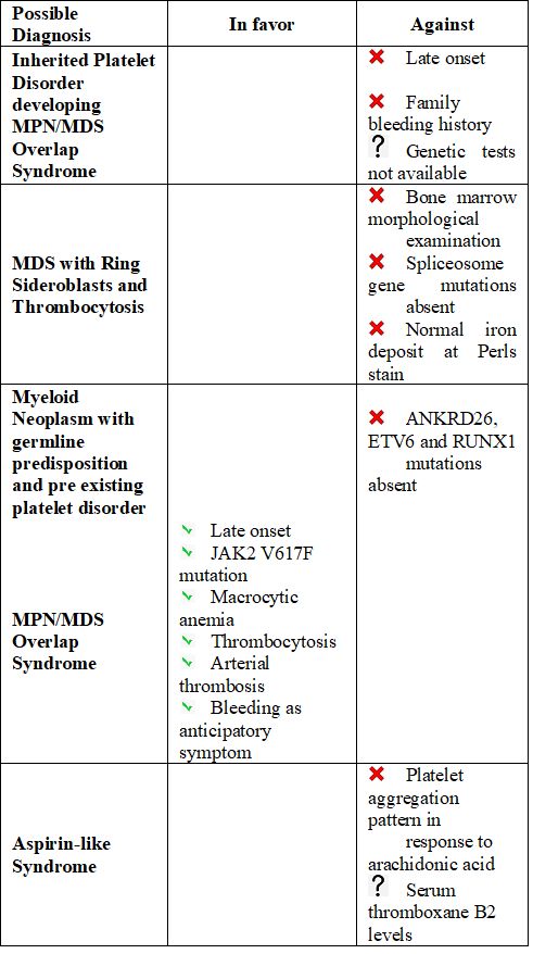
A possible MDS/MPN with ring sideroblasts and thrombocytosis (MDS/MPN-RS-T) was excluded, as ring sideroblasts were not appreciable and spliceosome gene mutations were not detected.
The patient was treated with hydroxyurea (4.5 g/week), resulting in a stable decrease of platelet count below 600 x 109/L. Hemoglobin levels significantly increased with rEPO 30.000 UI/week. An abdomen ultrasound exam showed the stability of the spleen six months after treatment had been started. A platelet aggregation test was repeated, confirming the same abnormalities shown previously. Antiplatelet prophylaxis has not yet been started despite the myeloproliferative aspect of the neoplasm and ischemic encephalopathy.
Certainly, the late onset of bleeding diathesis and the absence of hematologic neoplasms in family members made the hypothesis of a hereditary disease quite unlikely. We did not support the possibility of platelet disorders with a de novo germline predisposition to myeloid neoplasms as our patient was RUNX1/ ANKRD26/ETV6 negative; moreover, in this case, we would have expected a thrombocytopenic rather than a patient with thrombocytosis.
The reduced platelet response to so-called "weak agonists" (epinephrine) could suggest an "aspirin-like syndrome"[4] despite normal platelet aggregation in response to arachidonic acid. Serum thromboxane B2 levels would have been useful,[5] but they were not available for this patient.
We assumed the more likely hypothesis that platelet dysfunction could be the epiphenomenon of the hematological neoplasm that would have been diagnosed later.
Little information is available about platelet function and its role in many diseases, including MPN.[6] However, despite more infrequent than thrombosis, bleeding in MPN represents a more common phenomenon than in the general population[7] attributable 1) to a qualitative defect of von Willebrand Factor,[8] also in patients with controlled platelet count;[9] 2) a reduced expression or function of glycoproteins (GpIb or Gp IIb-IIIa);[10] 3) abnormalities in platelet granules.[11] As a consequence, a defective platelet aggregation pattern may be present in MPN, except for arachidonic acid and ristocetin as agonists.[10]
In conclusion, in this case, the hemorragic phenotype preceded the oncohematological diagnosis, appearing as the epiphenomenomen of the most prominent Myeloproliferative features in the setting of an MDS/MPN Overlap Syndrome.
The long-term outcome of this patient will definitely clarify the hematological diagnosis and the relationship between bleeding and oncohematological diagnosis.

References   


  1. Palomo L, Meggendorfer M, Hutter S, Twardziok S, Ademà V, Fuhrmann I, Fuster-Tormo F, Xicoy B, Zamora L, Acha P, Kerr CM, Kern W, Maciejewski JP, Solé F, Haferlach C, Haferlach T. Molecular landscape and clonal architecture of adult myelodysplastic/myeloproliferative neoplasms. Blood. 2020;136(16):1851-62. https://doi.org/10.1182/blood.2019004229 PMid:32573691 PMCid:PMC764560
  2. Visconte V, O Nakashima M, J Rogers H. Mutations in Splicing Factor Genes in Myeloid Malignancies: Significance and Impact on Clinical Features. Cancers (Basel). 2019;11(12):1844. https://doi.org/10.3390/cancers11121844 PMid:31766606 PMCid:PMC6966670
  3. Arber DA, Orazi A, Hasserjian R, Thiele J, Borowitz MJ, Le Beau MM, Bloomfield CD, Cazzola M, Vardiman JW. The 2016 revision to the World Health Organization classification of myeloid neoplasms and acute leukemia. Blood. 2016; 128(3):462-63 https://doi.org/10.1182/blood-2016-03-643544 PMid:27069254
  4. Rolf N, Knoefler R, Bugert P, Gehrisch S, Siegert G, Kuhlisch E, Suttorp M. Clinical and laboratory phenotypes associated with the aspirin-like defect: a study in 17 unrelated families. Br J Haematol. 2009; 144(3):416-24. https://doi.org/10.1111/j.1365-2141.2008.07468.x PMid:19036102
  5. Dragani A, Brancati F, Pascale S, Mattoscio D, Rocca B. Clinical and laboratory phenotype associated with the aspirin-like defect. Br J Haematol. 2010;148(4):661-63. https://doi.org/10.1111/j.1365-2141.2009.07950.x PMid:19814736
  6. Repsold L, Joubert AM. Platelet Function, Role in Thrombosis, Inflammation, and Consequences in Chronic Myeloproliferative Disorders. Cells. 2021;10(11):3034. https://doi.org/10.3390/cells10113034 PMid:34831257 PMCid:PMC8616365
  7. Patrono C, Rocca B, De Stefano V. Platelet activation and inhibition in polycythemia vera and essential thrombocythemia. Blood. 2013;121(10):1701-11. https://doi.org/10.1182/blood-2012-10-429134 PMid:23335367
  8. Castaman G, Lattuada A, Ruggeri M, Tosetto A, Mannucci PM, Rodeghiero F. Platelet von Willebrand factor abnormalities in myeloproliferative syndromes. Am J Hematol. 1995; 49(4):289-93. https://doi.org/10.1002/ajh.2830490406 PMid:7639273
  9. Lancellotti S, Dragani A, Ranalli P, Petrucci G, Basso M, Tartaglione R, Rocca B, De Cristofaro R. Qualitative and quantitative modifications of von Willebrand factor in patients with essential thrombocythemia and controlled platelet count. J Thromb Haemost. 2015 Jul;13(7):1226-37. https://doi.org/10.1111/jth.12967 PMid:25876231
  10. Barbui T, Cortelazzo S, Viero P. Thrombohaemorrhagic complications in 101 cases of myeloproliferative disorders: relationship to platelet number and function. Eur J Cancer Clin Oncol. 1983;19(11):1593-99 https://doi.org/10.1016/0277-5379(83)90091-3 PMid:6580171
  11. Gerrard JM, McNicol A. Platelet storage pool deficiency, leukemia, and myelodysplastic syndromes. Leuk Lymphoma. 1992;8(4-5):277-81. https://doi.org/10.3109/10428199209051007 PMid:1290957